Home BrowseSearch Help Group

Pfam Domains mapped on to the structure: 3F6A

No. Chain ID Pfam ID Pfam Description Linkout - Pfam Linkout - CDD
1 A PF00293 NUDIX domain PF00293 PF00293

Conserved Domain Database Superfamily Annotations: 3F6A

No. PDB ID PSSM ID CDD Accession Superfamily Short Name Linkout - CDD
1 3F6A 212606 cl00447 Nudix_Hydrolase superfamily - -
Structural Details of PDB entry 3F6A

Structural Details of PDB entry 3F6A

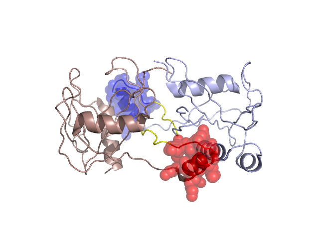
PDBid Chains Hinge Swapped Domain
3F6A A,B A:60-65,B:60-65;A:79-83,B:79-83 A:66-78,B:66-78

Swapped-domain interface residues and interactions:

Chains Residues
A 66, 67, 70, 71, 73, 74, 75, 76, 77, 78, 85, 86, 87, 88, 89, 135, 136, 137, 140,
B 66, 67, 70, 71, 73, 74, 75, 76, 77, 78, 85, 86, 87, 88, 135, 136, 137, 140,

Non-swapped-domain interface residues and interactions:

Chains Residues
A 1, 2, 3, 4, 5, 35, 37, 38, 39, 40, 41, 64, 79, 81, 82, 83, 84, 97, 99, 144, 150,
B 1, 2, 3, 4, 5, 35, 37, 38, 40, 41, 64, 79, 81, 82, 83, 84, 89, 97, 99, 144,

Swapped domains are represented using trasperent spheres.
Non-swapped part is represented using light color and cartoon representation.
Hinge region is shown in yellow color.

Mutations in critical regions:

Chains Hinge Domain swapped interface Non-swapped interface Swapped Domain
ANo mutationNo mutationNo mutationNo mutation
BNo mutationNo mutationNo mutationNo mutation

HIDE output:

Homologues found through HIDE algorithm

JMOL Visualization:

Jmol_logo            Jmol_logo            Jmol_logo

2D-plot:

B:3F6A A:3F6A

JOY Structural annotation for hinge hinge and swapped domain:

Hinge Swapped domain

JOY output:

ali file:3F6A.ali
atm file:3F6A.atm
cof file:3F6A.cof
hbd file:3F6A.hbd
html file:3F6A.html
pdb file:3F6A.pdb
ps file:3F6A.ps
psa file:3F6A.psa
rtf file:3F6A.rtf
sst file:3F6A.sst
tem file:3F6A.tem