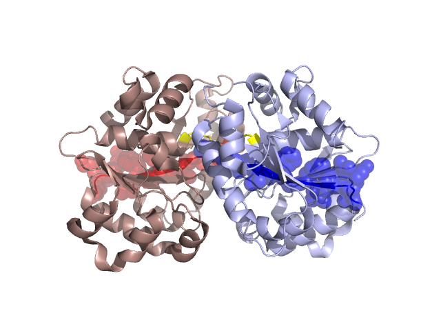
Structural Details of PDB entry 3EA0
Structural Details of PDB entry 3EA0
PDBid Chains Hinge Swapped Domain
3EA0
A,B
A:11-14,B:11-14
A:0-10,B:0-10
Swapped-domain interface residues and interactions:
Chains Residues
A
10 ,
B
10 ,
Non-swapped-domain interface residues and interactions:
Chains Residues
A
11 , 12 , 42 , 43 , 44 , 45 , 47 , 49 , 54 , 92 , 93 , 94 , 95 , 126 , 149 , 150 , 153 , 154 , 157 , 160 , 161 , 182 , 184 , 210 , 213 , 214 , 239 ,
B
11 , 12 , 42 , 43 , 44 , 45 , 47 , 49 , 92 , 93 , 94 , 126 , 127 , 149 , 150 , 153 , 154 , 157 , 160 , 161 , 164 , 183 , 184 , 185 , 210 , 211 , 213 , 214 ,