Home BrowseSearch Help Group

Pfam Domains mapped on to the structure: 3E8O

No. Chain ID Pfam ID Pfam Description Linkout - Pfam Linkout - CDD
1 A PF03992 Antibiotic biosynthesis monooxygenase PF03992 PF03992

Conserved Domain Database Superfamily Annotations: 3E8O

No. PDB ID PSSM ID CDD Accession Superfamily Short Name Linkout - CDD
1 3E8O 209125 cl10022 ABM superfamily - -
Structural Details of PDB entry 3E8O

Structural Details of PDB entry 3E8O

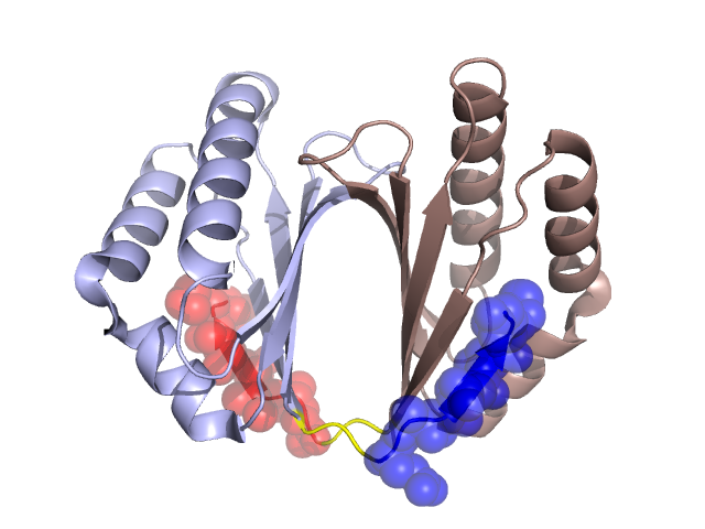
PDBid Chains Hinge Swapped Domain
3E8O A,B A:109-111,B:109-111 A:112-118,B:112-118

Swapped-domain interface residues and interactions:

Chains Residues
A 105, 106, 107, 108, 112, 113, 114, 115, 116, 118,
B 103, 104, 105, 106, 107, 108, 113, 114, 115, 116,

Non-swapped-domain interface residues and interactions:

Chains Residues
A 20, 22, 24, 36, 40, 47, 54, 56, 57, 58, 59, 60, 61, 62, 68, 70, 72, 76, 80, 83, 103, 104, 109, 110, 111,
B 20, 22, 24, 36, 40, 47, 54, 56, 57, 58, 59, 60, 61, 62, 68, 70, 72, 76, 83, 109, 110, 111,

Swapped domains are represented using trasperent spheres.
Non-swapped part is represented using light color and cartoon representation.
Hinge region is shown in yellow color.

Mutations in critical regions:

Chains Hinge Domain swapped interface Non-swapped interface Swapped Domain
ANo mutationNo mutationNo mutationNo mutation
BNo mutationNo mutationNo mutationNo mutation

HIDE output:

Homologues found through HIDE algorithm

JMOL Visualization:

Jmol_logo            Jmol_logo            Jmol_logo

2D-plot:

A:3E8O B:3E8O

JOY Structural annotation for hinge hinge and swapped domain:

Hinge Swapped domain

JOY output:

ali file:3E8O.ali
atm file:3E8O.atm
cof file:3E8O.cof
hbd file:3E8O.hbd
html file:3E8O.html
pdb file:3E8O.pdb
ps file:3E8O.ps
psa file:3E8O.psa
rtf file:3E8O.rtf
sst file:3E8O.sst
tem file:3E8O.tem