Home BrowseSearch Help Group

Pfam Domains mapped on to the structure: 3DWI

No. Chain ID Pfam ID Pfam Description Linkout - Pfam Linkout - CDD
1 A PF00291 Pyridoxal-phosphate dependent enzyme PF00291 PF00291

Conserved Domain Database Superfamily Annotations: 3DWI

No. PDB ID PSSM ID CDD Accession Superfamily Short Name Linkout - CDD
1 3DWI 107204 cd01561 CBS_like - cl00342
2 3DWI 212605 cl00342 Trp-synth-beta_II superfamily - -
3 3DWI 212605 cl00342 Trp-synth-beta_II superfamily - -
Structural Details of PDB entry 3DWI

Structural Details of PDB entry 3DWI

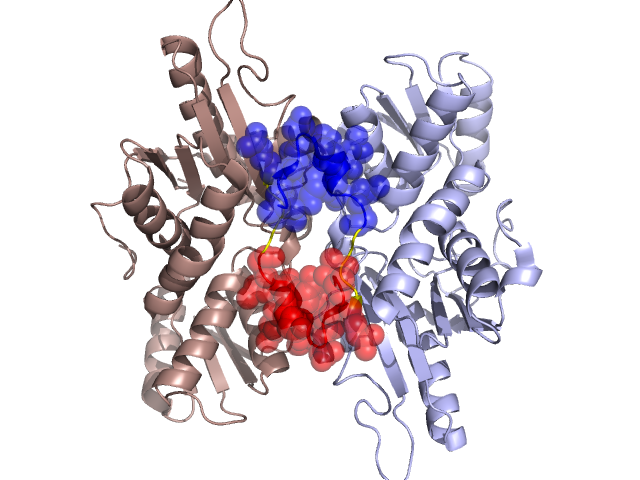
PDBid Chains Hinge Swapped Domain
3DWI A,B A:13-16,B:13-16 A:1-12,B:1-12

Swapped-domain interface residues and interactions:

Chains Residues
A 1, 2, 3, 4, 5, 6, 7, 10, 15, 16, 17, 18, 172,
B 1, 2, 3, 4, 5, 6, 7, 10, 13, 15, 16, 17, 18, 172,

Non-swapped-domain interface residues and interactions:

Chains Residues
A 13, 20, 21, 26, 38, 44, 46, 47, 48, 84, 88, 91, 92, 111, 112, 115, 116, 117, 171, 175, 255, 256, 257, 258, 259, 260, 261, 298, 299, 301, 302, 306, 307, 314,
B 20, 21, 38, 44, 46, 47, 48, 88, 91, 92, 94, 111, 112, 115, 116, 117, 175, 255, 256, 257, 258, 259, 260, 261, 298, 299, 301, 302, 306, 307,

Swapped domains are represented using trasperent spheres.
Non-swapped part is represented using light color and cartoon representation.
Hinge region is shown in yellow color.

Mutations in critical regions:

Chains Hinge Domain swapped interface Non-swapped interface Swapped Domain
ANo mutationNo mutationNo mutationNo mutation
BNo mutationNo mutationNo mutationNo mutation

HIDE output:

Homologues found through HIDE algorithm

JMOL Visualization:

Jmol_logo            Jmol_logo            Jmol_logo

2D-plot:

A:3DWI B:3DWI

JOY Structural annotation for hinge hinge and swapped domain:

Hinge Swapped domain

JOY output:

ali file:3DWI.ali
atm file:3DWI.atm
cof file:3DWI.cof
hbd file:3DWI.hbd
html file:3DWI.html
pdb file:3DWI.pdb
ps file:3DWI.ps
psa file:3DWI.psa
rtf file:3DWI.rtf
sst file:3DWI.sst
tem file:3DWI.tem