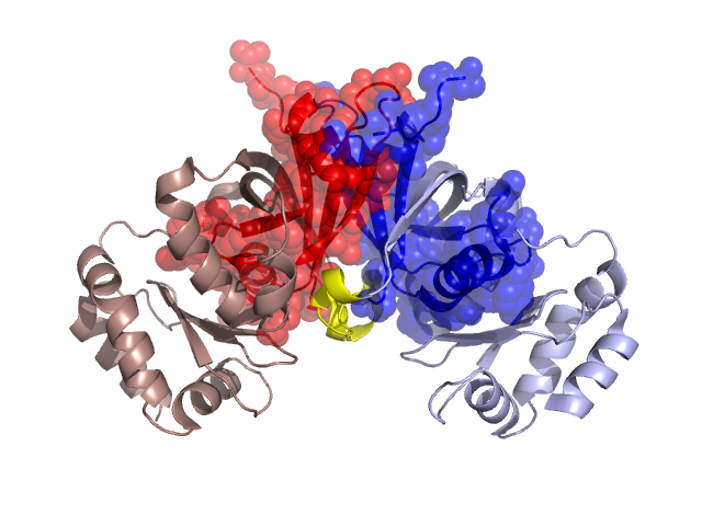
Structural Details of PDB entry 3DNH
Structural Details of PDB entry 3DNH
PDBid Chains Hinge Swapped Domain
3DNH
A,B
A:94-101,B:94-101
A:4-93,B:5-93
Swapped-domain interface residues and interactions:
Chains Residues
A
6 , 8 , 9 , 10 , 12 , 39 , 41 , 43 , 45 , 46 , 48 , 49 , 50 , 51 , 52 , 53 , 54 , 55 , 56 , 57 , 58 , 70 , 73 , 74 , 75 , 76 , 77 , 79 , 86 , 88 , 90 , 98 , 99 , 100 , 106 , 108 ,
B
6 , 8 , 9 , 10 , 12 , 38 , 39 , 41 , 43 , 45 , 46 , 48 , 49 , 50 , 51 , 52 , 53 , 54 , 55 , 56 , 57 , 58 , 70 , 73 , 74 , 75 , 76 , 79 , 86 , 88 , 90 , 94 , 95 , 98 , 99 , 100 , 104 , 106 , 108 ,
Non-swapped-domain interface residues and interactions:
Chains Residues
A
94 , 95 , 104 , 130 , 132 , 136 , 153 , 155 , 254 ,
B
97 , 101 , 136 , 153 , 155 ,