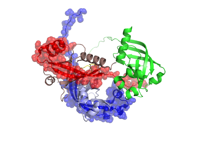
Structural Details of PDB entry 3DMB
Structural Details of PDB entry 3DMB
PDBid Chains Hinge Swapped Domain
3DMB
A,B
A:75-78,B:75-78
A:79-145,B:79-145
Swapped-domain interface residues and interactions:
Chains Residues
A
33 , 80 , 82 , 102 , 103 , 106 , 126 , 128 , 130 , 131 , 135 , 136 , 137 , 140 , 145 ,
B
32 , 33 , 35 , 80 , 82 , 102 , 103 , 106 , 107 , 128 , 130 , 137 , 145 ,
Non-swapped-domain interface residues and interactions:
Chains Residues
A
0 , 2 , 20 , 26 , 27 , 28 , 31 , 34 , 35 , 69 , 71 , 73 , 75 , 76 , 77 ,
B
18 , 20 , 26 , 30 , 31 , 34 , 69 , 71 , 73 , 75 , 76 , 77 ,
C
16 , 20 , 35 , 101 , 102 , 103 ,