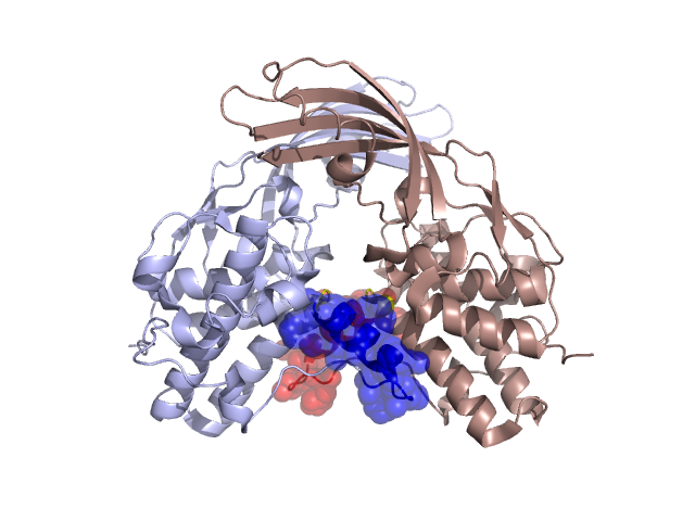
Structural Details of PDB entry 3DA3
Structural Details of PDB entry 3DA3
PDBid Chains Hinge Swapped Domain
3DA3
A,B
A:24-28,B:24-28;A:46-48,B:46-48
A:29-45,B:29-45
Swapped-domain interface residues and interactions:
Chains Residues
A
28 , 29 , 30 , 31 , 35 , 36 , 39 , 40 , 43 , 44 , 48 , 49 , 53 , 56 , 57 , 60 , 238 ,
B
29 , 30 , 31 , 35 , 36 , 39 , 40 , 43 , 48 , 49 , 53 , 56 , 57 , 60 , 238 ,
Non-swapped-domain interface residues and interactions:
Chains Residues
A
26 , 47 , 52 , 95 , 154 , 190 , 245 , 264 , 266 , 271 ,
B
26 , 28 , 47 , 52 , 95 , 154 , 190 , 245 , 262 , 264 ,