Pfam Domains mapped on to the structure: 3D7I
No.
Chain ID
Pfam ID
Pfam Description
Linkout - Pfam
Linkout - CDD
1
A
PF02627
Carboxymuconolactone decarboxylase family
PF02627
PF02627
Conserved Domain Database Superfamily Annotations: 3D7I
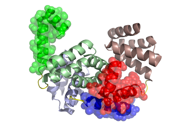
Structural Details of PDB entry 3D7I
Structural Details of PDB entry 3D7I
PDBid Chains Hinge Swapped Domain
3D7I
A,C,B
A:35-38,C:35-38,B:35-38
A:8-34,C:8-34,B:8-34
Swapped-domain interface residues and interactions:
Chains Residues
A
9 , 10 , 13 , 14 , 18 , 20 , 21 , 24 , 25 , 27 , 28 , 31 , 32 , 51 ,
B
28 , 29 , 31 , 32 , 33 , 51 , 53 , 83 , 84 , 87 , 95 , 96 , 99 ,
C
17 , 18 , 20 , 21 , 24 , 27 , 28 , 29 , 31 , 32 , 33 , 34 , 51 , 83 , 84 , 87 ,
Non-swapped-domain interface residues and interactions:
Chains Residues
A
36 , 54 , 55 , 56 , 58 , 59 , 104 ,
B
35 , 36 , 37 , 38 , 42 , 43 , 45 , 46 , 47 , 49 , 50 , 54 , 55 , 76 , 77 , 79 , 80 , 81 , 86 , 88 , 89 , 92 , 94 , 102 , 104 ,
C
35 , 36 , 37 , 38 , 42 , 43 , 45 , 46 , 47 , 49 , 50 , 53 , 54 , 76 , 77 , 80 , 81 , 86 , 88 , 89 ,
Mutations in critical regions:
Chains
Hinge
Domain swapped interface Non-swapped interface Swapped Domain
A No mutation No mutation No mutation ALA(15)LYS, ALA(16)GLU, ALA(17)LYS, C No mutation ALA(17)LYS, No mutation ALA(15)LYS, ALA(16)GLU, ALA(17)LYS, B No mutation No mutation No mutation ALA(15)LYS, ALA(16)GLU, ALA(17)LYS,
HIDE output:
JMOL Visualization:
2D-plot:
JOY Structural annotation for hinge hinge and swapped domain:
JOY output: