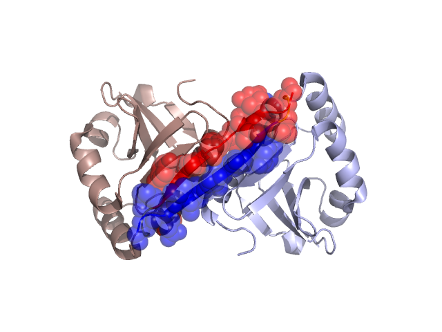
Pfam Domains mapped on to the structure: 3D5P No. Chain ID Pfam ID Pfam Description Linkout - Pfam Linkout - CDD 1 A PF09346 SMI1 / KNR4 family (SUKH-1) PF09346 PF09346 Conserved Domain Database Superfamily Annotations: 3D5P No. PDB ID PSSM ID CDD Accession Superfamily Short Name Linkout - CDD 1 3D5P 207472 cl01747 SMI1_KNR4 superfamily - - Structural Details of PDB entry 3D5P Structural Details of PDB entry 3D5P PDBid Chains Hinge Swapped Domain 3D5P A,B A:11-13,B:11-13 A:0-10,B:0-10 Swapped-domain interface residues and interactions: Chains Residues A 0, 2, 3, 4, 5, 6, 7, 8, 9, 10, 12, 36, 126, B 0, 2, 3, 4, 5, 6, 7, 8, 9, 10, 12, 36, 126, Non-swapped-domain interface residues and interactions: Chains Residues A 33, 37, 40, 48, 119, 120, 122, 123, 124, 127, 132, B 33, 37, 40, 48, 119, 120, 122, 123, 124, 127, Swapped domains are represented using trasperent spheres. Non-swapped part is represented using light color and cartoon representation. Hinge region is shown in yellow color. Mutations in critical regions: Chains Hinge Domain swapped interface Non-swapped interface Swapped Domain ANo mutationNo mutationNo mutationNo mutation BNo mutationNo mutationNo mutationNo mutation HIDE output: Homologues found through HIDE algorithm JMOL Visualization: 2D-plot: A:3D5P B:3D5P JOY Structural annotation for hinge hinge and swapped domain: Hinge Swapped domain JOY output: ali file:3D5P.ali atm file:3D5P.atm cof file:3D5P.cof hbd file:3D5P.hbd html file:3D5P.html pdb file:3D5P.pdb ps file:3D5P.ps psa file:3D5P.psa rtf file:3D5P.rtf sst file:3D5P.sst tem file:3D5P.tem