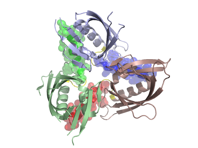
Pfam Domains mapped on to the structure: 3CM1
No.
Chain ID
Pfam ID
Pfam Description
Linkout - Pfam
Linkout - CDD
1
A
PF04686
Streptomyces sporulation and cell division protein, SsgA
PF04686
PF04686
Conserved Domain Database Superfamily Annotations: 3CM1
Structural Details of PDB entry 3CM1
Structural Details of PDB entry 3CM1
PDBid Chains Hinge Swapped Domain
3CM1
A,C,B
A:118-121,C:118-121,B:118-121
A:122-137,C:122-137,B:122-137
Swapped-domain interface residues and interactions:
Chains Residues
A
62 , 63 , 65 , 66 , 108 , 120 , 121 , 124 , 127 , 128 , 130 , 131 , 133 , 134 , 135 , 137 ,
B
62 , 63 , 65 , 66 , 108 , 120 , 121 , 124 , 127 , 128 , 130 , 131 , 133 , 134 , 135 , 137 ,
C
59 , 62 , 63 , 66 , 108 , 120 , 121 , 124 , 127 , 128 , 130 , 131 , 133 , 134 , 135 ,
Non-swapped-domain interface residues and interactions:
Chains Residues
A
33 , 35 , 36 , 38 , 41 , 53 , 55 , 59 , 67 , 68 , 69 , 70 , 71 , 72 , 85 , 104 , 105 , 112 , 115 ,
B
33 , 35 , 36 , 38 , 41 , 53 , 55 , 59 , 67 , 68 , 69 , 70 , 71 , 75 , 85 , 104 , 112 , 115 ,
C
33 , 35 , 36 , 38 , 41 , 53 , 55 , 65 , 67 , 68 , 69 , 70 , 71 , 72 , 75 , 85 , 104 , 112 , 115 ,
Mutations in critical regions:
Chains
Hinge
Domain swapped interface Non-swapped interface Swapped Domain
A No mutation No mutation No mutation No mutation C No mutation No mutation No mutation No mutation B No mutation No mutation No mutation No mutation
HIDE output:
JMOL Visualization:
2D-plot:
JOY Structural annotation for hinge hinge and swapped domain:
JOY output: