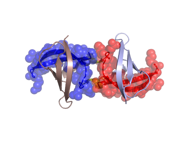
Structural Details of PDB entry 3CAM
Structural Details of PDB entry 3CAM
PDBid Chains Hinge Swapped Domain
3CAM
A,B
A:36-39,B:36-39
A:40-67,B:40-67
Swapped-domain interface residues and interactions:
Chains Residues
A
2 , 3 , 4 , 5 , 6 , 7 , 8 , 9 , 27 , 28 , 29 , 31 , 33 , 34 , 35 , 38 , 39 , 40 , 41 , 42 , 43 , 44 , 45 , 46 , 47 , 48 , 49 , 50 , 52 , 57 , 59 , 60 , 61 , 62 , 63 , 64 , 65 , 66 , 67 ,
B
2 , 3 , 4 , 5 , 6 , 7 , 8 , 9 , 27 , 28 , 29 , 31 , 33 , 34 , 35 , 38 , 39 , 40 , 41 , 42 , 43 , 44 , 45 , 46 , 47 , 48 , 49 , 50 , 52 , 57 , 59 , 60 , 61 , 62 , 63 , 64 , 65 , 66 ,
Non-swapped-domain interface residues and interactions: