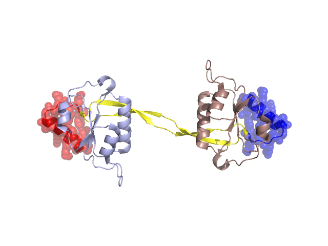
Pfam Domains mapped on to the structure: 3CAE
No.
Chain ID
Pfam ID
Pfam Description
Linkout - Pfam
Linkout - CDD
1
A
PF05367
Phage endonuclease I
PF05367
PF05367
Conserved Domain Database Superfamily Annotations: 3CAE
Structural Details of PDB entry 3CAE
Structural Details of PDB entry 3CAE
PDBid Chains Hinge Swapped Domain
3CAE
A,B
A:37-59,B:37-59
A:17-36,B:17-36
Swapped-domain interface residues and interactions:
Chains Residues
A
17 , 19 , 20 , 22 , 23 , 26 , 27 , 30 , 32 , 33 , 34 , 35 , 36 , 58 , 59 , 60 , 61 , 62 , 69 , 98 , 126 , 127 , 128 ,
B
17 , 19 , 20 , 22 , 23 , 26 , 27 , 30 , 32 , 33 , 34 , 35 , 36 , 58 , 59 , 60 , 61 , 62 , 69 , 98 , 126 , 127 , 128 ,
Non-swapped-domain interface residues and interactions:
Chains Residues
A
37 , 38 , 39 , 40 , 41 , 42 , 43 , 44 , 45 , 46 , 47 , 48 , 49 , 50 , 51 , 52 , 53 , 54 , 55 , 56 , 57 , 71 , 80 , 83 , 84 , 87 , 88 , 91 , 96 , 130 , 133 , 142 , 145 , 146 , 147 , 148 ,
B
37 , 38 , 39 , 40 , 41 , 42 , 43 , 44 , 45 , 46 , 47 , 48 , 49 , 50 , 51 , 52 , 53 , 54 , 55 , 56 , 57 , 66 , 71 , 80 , 83 , 84 , 87 , 88 , 91 , 96 , 130 , 133 , 142 , 144 , 145 , 146 , 147 ,
Mutations in critical regions:
Chains
Hinge
Domain swapped interface Non-swapped interface Swapped Domain
A SER(44)-, ASN(45)-, ASN(46)-, GLN(47)-, GLN(48)-, ASN(49)-, TYR(50)-, SER(51)-, SER(52)-, MET(17)-, SER(44)-, ASN(45)-, ASN(46)-, GLN(47)-, GLN(48)-, ASN(49)-, TYR(50)-, SER(51)-, SER(52)-, MET(17)-, B SER(44)-, ASN(45)-, ASN(46)-, GLN(47)-, GLN(48)-, ASN(49)-, TYR(50)-, SER(51)-, SER(52)-, MET(17)-, SER(44)-, ASN(45)-, ASN(46)-, GLN(47)-, GLN(48)-, ASN(49)-, TYR(50)-, SER(51)-, SER(52)-, MET(17)-,
HIDE output:
JMOL Visualization:
2D-plot:
JOY Structural annotation for hinge hinge and swapped domain:
JOY output: