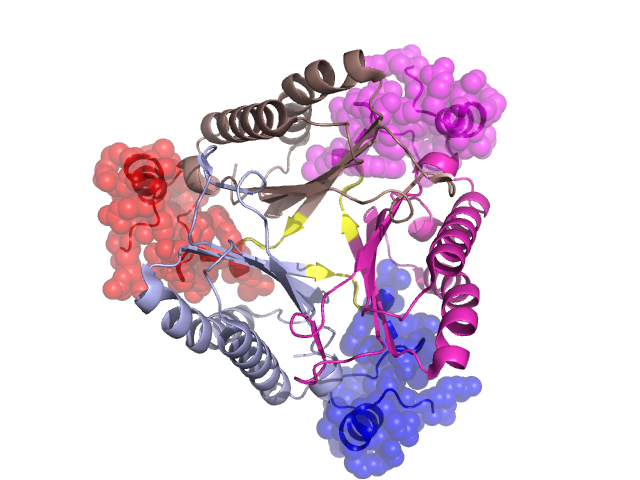
Structural Details of PDB entry 3C6V
Structural Details of PDB entry 3C6V
PDBid Chains Hinge Swapped Domain
3C6V
A,D,C,B
A:107-110,D:107-110,C:107-110,B:107-110
A:111-140,D:111-140,C:111-140,B:111-140
Swapped-domain interface residues and interactions:
Chains Residues
A
87 , 100 , 101 , 102 , 103 , 104 , 105 , 111 , 112 , 113 , 114 , 115 , 116 , 117 , 140 ,
B
87 , 100 , 101 , 102 , 103 , 104 , 105 , 111 , 112 , 113 , 114 , 115 , 116 , 117 , 140 ,
C
87 , 100 , 101 , 102 , 103 , 104 , 105 , 111 , 112 , 113 , 114 , 115 , 116 , 117 ,
Non-swapped-domain interface residues and interactions:
Chains Residues
A
3 , 5 , 7 , 15 , 18 , 19 , 22 , 23 , 26 , 36 , 37 , 39 , 40 , 41 , 42 , 43 , 44 , 46 , 49 , 50 , 51 , 52 , 53 , 54 , 55 , 63 , 65 , 67 , 79 , 83 , 106 , 107 , 108 , 109 ,
B
3 , 3 , 5 , 7 , 15 , 18 , 19 , 22 , 23 , 26 , 36 , 37 , 39 , 40 , 41 , 42 , 43 , 44 , 46 , 49 , 50 , 51 , 52 , 53 , 54 , 55 , 57 , 63 , 65 , 67 , 71 , 74 , 79 , 83 , 106 , 107 , 108 , 109 ,
C
3 , 5 , 7 , 15 , 18 , 19 , 22 , 23 , 26 , 36 , 37 , 39 , 40 , 41 , 42 , 43 , 44 , 46 , 49 , 50 , 51 , 52 , 53 , 54 , 55 , 56 , 63 , 65 , 67 , 69 , 79 , 83 , 106 , 107 , 108 , 109 ,
Mutations in critical regions:
Chains
Hinge
Domain swapped interface Non-swapped interface Swapped Domain
A No mutation No mutation No mutation No mutation D No mutation No mutation No mutation No mutation C No mutation No mutation No mutation No mutation B No mutation No mutation No mutation No mutation
HIDE output:
JMOL Visualization:
2D-plot:
JOY Structural annotation for hinge hinge and swapped domain:
JOY output: