Pfam Domains mapped on to the structure: 3C6F
No.
Chain ID
Pfam ID
Pfam Description
Linkout - Pfam
Linkout - CDD
1
A
PF04239
Protein of unknown function (DUF421)
PF04239
PF04239
Conserved Domain Database Superfamily Annotations: 3C6F
No.
PDB ID
PSSM ID
CDD Accession
Superfamily Short Name
Linkout - CDD
1
3C6F
207274
DUF421
superfamily
N -
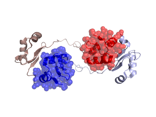
Structural Details of PDB entry 3C6F
Structural Details of PDB entry 3C6F
PDBid Chains Hinge Swapped Domain
3C6F
A,D,C,B
A:54-58,D:54-58,C:54-58,B:54-58
A:94-153,D:94-153,C:94-153,B:94-153
Swapped-domain interface residues and interactions:
Chains Residues
A
101 , 103 , 105 , 106 , 115 , 116 , 119 , 128 , 129 , 130 , 131 , 132 , 152 , 170 , 228 , 229 ,
C
101 , 102 , 103 , 105 , 106 , 115 , 116 , 119 , 128 , 129 , 130 , 131 , 170 , 228 ,
Non-swapped-domain interface residues and interactions:
Chains Residues
A
169 , 172 , 175 , 176 , 210 , 211 , 213 , 215 , 221 , 223 , 227 , 230 ,
C
169 , 172 , 175 , 176 , 210 , 211 , 213 , 215 , 218 , 221 , 223 , 229 ,
Mutations in critical regions:
Chains
Hinge
Domain swapped interface Non-swapped interface Swapped Domain
A No mutation No mutation No mutation MET(121)LEU, MET(138)LEU, D No mutation No mutation No mutation MET(121)LEU, MET(138)LEU, C No mutation No mutation No mutation MET(121)LEU, MET(138)LEU, B No mutation No mutation No mutation MET(121)LEU, MET(138)LEU,
HIDE output:
JMOL Visualization:
2D-plot:
JOY Structural annotation for hinge hinge and swapped domain:
JOY output: