Home BrowseSearch Help Group

Pfam Domains mapped on to the structure: 3C3M

No. Chain ID Pfam ID Pfam Description Linkout - Pfam Linkout - CDD
1 A PF00072 Response regulator receiver domain PF00072 PF00072

Gene Ontology Annotations: 3C3M

No. GO ID GO Description Linkout - AmiGO
1 GO:0006355 regulation of transcription, DNA-dependent GO:0006355

Conserved Domain Database Superfamily Annotations: 3C3M

No. PDB ID PSSM ID CDD Accession Superfamily Short Name Linkout - CDD
1 3C3M 29071 cd00156 REC - cl09944
2 3C3M 209106 cl09944 REC superfamily - -
3 3C3M 209106 cl09944 REC superfamily - -
Structural Details of PDB entry 3C3M

Structural Details of PDB entry 3C3M

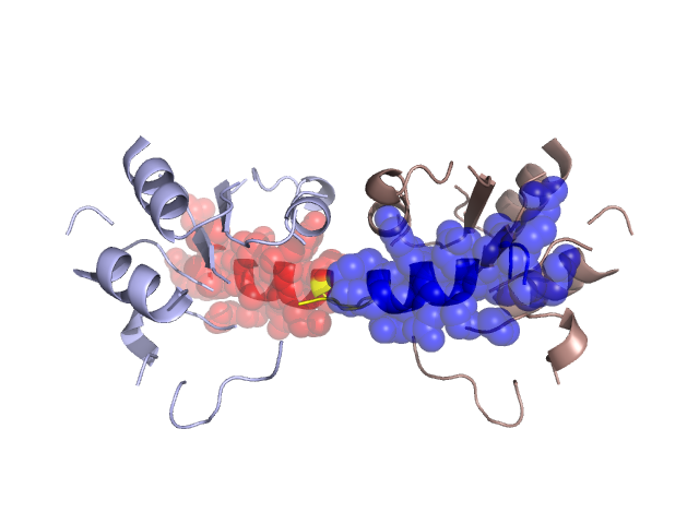
PDBid Chains Hinge Swapped Domain
3C3M A,B A:104-108,B:104-108 A:109-123,B:109-123

Swapped-domain interface residues and interactions:

Chains Residues
A 111, 112, 113, 115, 116, 117, 119, 120, 121, 123,
B 111, 112, 113, 115, 116, 117, 119, 120, 121,

Non-swapped-domain interface residues and interactions:

Chains Residues
A 3, 5, 12, 16, 17, 18, 20, 22, 24, 25, 27, 48, 49, 78, 80, 100, 101, 103, 104, 106, 108,
B 3, 5, 12, 16, 17, 18, 20, 22, 24, 25, 27, 48, 49, 78, 80, 100, 101, 103, 104, 106, 108,

Swapped domains are represented using trasperent spheres.
Non-swapped part is represented using light color and cartoon representation.
Hinge region is shown in yellow color.

Mutations in critical regions:

Chains Hinge Domain swapped interface Non-swapped interface Swapped Domain
ANo mutationNo mutationNo mutationNo mutation
BNo mutationNo mutationNo mutationNo mutation

HIDE output:

Homologues found through HIDE algorithm

JMOL Visualization:

Jmol_logo            Jmol_logo            Jmol_logo

2D-plot:

A:3C3M B:3C3M

JOY Structural annotation for hinge hinge and swapped domain:

Hinge Swapped domain

JOY output:

ali file:3C3M.ali
atm file:3C3M.atm
cof file:3C3M.cof
hbd file:3C3M.hbd
html file:3C3M.html
pdb file:3C3M.pdb
ps file:3C3M.ps
psa file:3C3M.psa
rtf file:3C3M.rtf
sst file:3C3M.sst
tem file:3C3M.tem