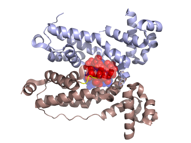
Structural Details of PDB entry 3C2B
Structural Details of PDB entry 3C2B
PDBid Chains Hinge Swapped Domain
3C2B
A,B
A:170-172,B:170-172;A:184-186,B:184-186
A:173-183,B:173-183
Swapped-domain interface residues and interactions:
Chains Residues
A
115 , 118 , 126 , 130 , 168 , 173 , 174 , 175 , 176 , 177 , 179 , 180 , 181 , 182 ,
B
115 , 118 , 120 , 122 , 126 , 130 , 168 , 173 , 175 , 176 , 177 , 179 , 180 , 181 , 182 ,
Non-swapped-domain interface residues and interactions:
Chains Residues
A
27 , 28 , 29 , 30 , 31 , 32 , 105 , 111 , 112 , 116 , 119 , 129 , 134 , 161 , 164 , 165 , 167 , 169 , 172 , 191 , 194 , 198 , 201 , 202 , 214 ,
B
111 , 112 , 116 , 117 , 119 , 121 , 129 , 134 , 161 , 164 , 165 , 167 , 169 , 172 , 185 , 191 , 194 , 198 , 201 , 202 ,