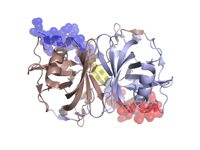
Pfam Domains mapped on to the structure: 3BNK
No.
Chain ID
Pfam ID
Pfam Description
Linkout - Pfam
Linkout - CDD
1
A
PF01613
Flavin reductase like domain
PF01613
PF01613
Conserved Domain Database Superfamily Annotations: 3BNK
Structural Details of PDB entry 3BNK
Structural Details of PDB entry 3BNK
PDBid Chains Hinge Swapped Domain
3BNK
A,B
A:171-174,B:171-174
A:175-187,B:175-188
Swapped-domain interface residues and interactions:
Chains Residues
A
78 , 79 , 81 , 82 , 84 , 85 , 167 , 168 , 169 , 170 , 171 , 175 , 176 , 177 , 178 , 179 , 180 , 182 , 183 , 184 , 185 , 186 , 187 ,
B
78 , 79 , 81 , 82 , 84 , 85 , 167 , 168 , 169 , 170 , 171 , 175 , 176 , 177 , 178 , 179 , 180 , 182 , 183 , 184 , 185 , 186 , 187 ,
Non-swapped-domain interface residues and interactions:
Chains Residues
A
2 , 3 , 4 , 5 , 6 , 7 , 9 , 10 , 11 , 12 , 13 , 14 , 15 , 17 , 36 , 37 , 38 , 39 , 40 , 41 , 42 , 43 , 44 , 45 , 46 , 47 , 50 , 52 , 71 , 77 , 86 , 100 , 103 , 111 , 115 , 117 , 123 , 125 , 127 , 128 , 129 , 130 , 132 , 134 , 137 , 138 , 139 , 140 , 141 , 142 , 143 , 144 , 147 , 150 , 151 , 152 , 154 , 155 , 157 , 159 , 161 , 163 , 164 , 165 , 166 , 172 , 173 , 174 ,
B
2 , 3 , 4 , 5 , 6 , 7 , 9 , 10 , 11 , 13 , 14 , 15 , 17 , 36 , 37 , 38 , 39 , 40 , 41 , 42 , 43 , 44 , 45 , 46 , 47 , 52 , 71 , 77 , 86 , 100 , 103 , 111 , 115 , 117 , 123 , 125 , 127 , 128 , 129 , 130 , 132 , 134 , 137 , 138 , 139 , 140 , 141 , 142 , 143 , 144 , 147 , 150 , 151 , 152 , 154 , 155 , 157 , 159 , 161 , 163 , 164 , 165 , 172 , 173 , 174 ,
Mutations in critical regions:
Chains
Hinge
Domain swapped interface Non-swapped interface Swapped Domain
A No mutation No mutation No mutation No mutation B No mutation No mutation No mutation No mutation
HIDE output:
JMOL Visualization:
2D-plot:
JOY Structural annotation for hinge hinge and swapped domain:
JOY output: