Pfam Domains mapped on to the structure: 3BIO
Gene Ontology Annotations: 3BIO
Conserved Domain Database Superfamily Annotations: 3BIO
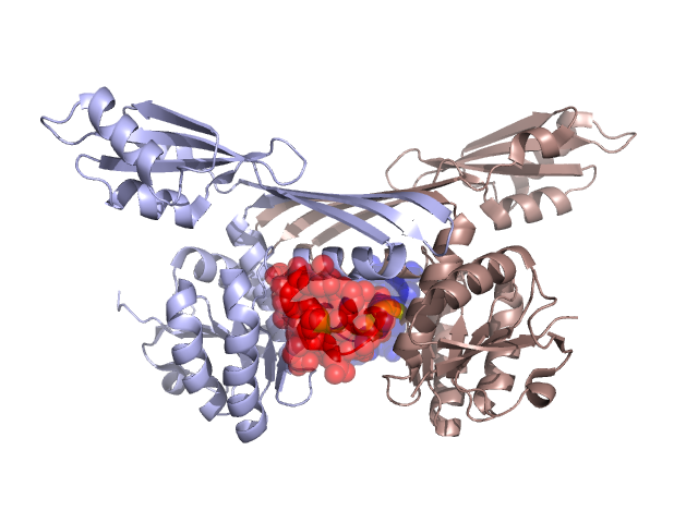
Structural Details of PDB entry 3BIO
Structural Details of PDB entry 3BIO
PDBid Chains Hinge Swapped Domain
3BIO
A,B
A:280-284,B:280-284
A:285-301,B:285-301
Swapped-domain interface residues and interactions:
Chains Residues
A
100 , 129 , 130 , 132 , 133 , 134 , 137 , 281 , 282 , 285 , 286 , 288 , 289 , 293 , 294 , 296 , 297 , 298 , 300 ,
B
100 , 129 , 130 , 132 , 133 , 134 , 137 , 281 , 282 , 285 , 286 , 288 , 289 , 293 , 294 , 296 , 297 , 298 , 300 ,
Non-swapped-domain interface residues and interactions:
Chains Residues
A
23 , 24 , 27 , 96 , 99 , 103 , 127 , 131 , 136 , 138 , 140 , 141 , 239 , 240 , 241 , 242 , 245 , 246 , 248 , 249 , 250 , 252 , 253 , 254 , 257 , 258 , 261 , 262 , 265 , 279 , 284 ,
B
23 , 24 , 27 , 96 , 99 , 103 , 127 , 131 , 136 , 138 , 140 , 141 , 239 , 240 , 241 , 242 , 245 , 246 , 248 , 249 , 250 , 252 , 253 , 254 , 257 , 258 , 261 , 262 , 265 , 279 , 284 ,
Mutations in critical regions:
Chains
Hinge
Domain swapped interface Non-swapped interface Swapped Domain
A No mutation No mutation No mutation No mutation B No mutation No mutation No mutation No mutation
HIDE output:
JMOL Visualization:
2D-plot:
JOY Structural annotation for hinge hinge and swapped domain:
JOY output: