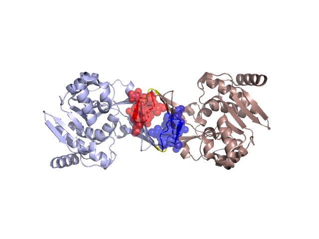
Pfam Domains mapped on to the structure: 3BF5 No. Chain ID Pfam ID Pfam Description Linkout - Pfam Linkout - CDD 1 A PF00294 pfkB family carbohydrate kinase PF00294 PF00294 Conserved Domain Database Superfamily Annotations: 3BF5 No. PDB ID PSSM ID CDD Accession Superfamily Short Name Linkout - CDD 1 3BF5 29364 cd01942 ribokinase_group_A - cl00192 2 3BF5 212177 cl00192 ribokinase_pfkB_like superfamily - - 3 3BF5 212177 cl00192 ribokinase_pfkB_like superfamily - - Structural Details of PDB entry 3BF5 Structural Details of PDB entry 3BF5 PDBid Chains Hinge Swapped Domain 3BF5 A,B A:17-19,B:17-19;A:29-31,B:29-31 A:20-28,B:20-28 Swapped-domain interface residues and interactions: Chains Residues A 21, 22, 23, 24, 25, 26, 27, 28, 101, 102, 103, 104, 105, 107, B 21, 22, 23, 24, 25, 26, 27, 28, 101, 102, 103, 104, 105, Non-swapped-domain interface residues and interactions: Chains Residues A 14, 16, 29, 32, 89, 94, 96, 108, 283, B 14, 16, 29, 32, 89, 94, 96, 107, 108, Swapped domains are represented using trasperent spheres. Non-swapped part is reprented using light color and cartoon reprentation. Hinge region is shown in yellow color. Mutations in critical regions: Chains Hinge Domain swapped interface Non-swapped interface Swapped Domain ANo mutationNo mutationNo mutationNo mutation BNo mutationNo mutationNo mutationNo mutation BNo mutationNo mutationNo mutationNo mutation HIDE output: Homologues found through HIDE algorithm JMOL Visualization: 2D-plot: B:3BF5 3:3BF5 A:3BF5 JOY Structural annotation for hinge hinge and swapped domain: Hinge Swapped domain JOY output: ali file:3BF5.ali atm file:3BF5.atm cof file:3BF5.cof hbd file:3BF5.hbd html file:3BF5.html pdb file:3BF5.pdb ps file:3BF5.ps psa file:3BF5.psa rtf file:3BF5.rtf sst file:3BF5.sst tem file:3BF5.tem