Home BrowseSearch Help Group

Pfam Domains mapped on to the structure: 3BEY

No. Chain ID Pfam ID Pfam Description Linkout - Pfam Linkout - CDD
1 A PF02627 Carboxymuconolactone decarboxylase family PF02627 PF02627

Gene Ontology Annotations: 3BEY

No. GO ID GO Description Linkout - AmiGO
1 GO:0055114 oxidation-reduction process GO:0055114

Conserved Domain Database Superfamily Annotations: 3BEY

No. PDB ID PSSM ID CDD Accession Superfamily Short Name Linkout - CDD
1 3BEY 212222 CMD superfamily C -
Structural Details of PDB entry 3BEY

Structural Details of PDB entry 3BEY

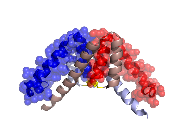
PDBid Chains Hinge Swapped Domain
3BEY A,C A:48-49,C:48-49 A:7-47,C:4-47

Swapped-domain interface residues and interactions:

Chains Residues
A 7, 8, 11, 19, 22, 23, 26, 29, 31, 32, 35, 36, 37, 39, 40, 41, 43, 44, 45, 47, 48, 70, 76, 77, 80, 81, 88, 90, 91,
C 4, 5, 7, 8, 11, 19, 22, 23, 26, 29, 31, 32, 35, 36, 37, 39, 40, 41, 43, 44, 45, 47, 48, 70, 73, 76, 77, 78, 80, 81, 90, 91,

Non-swapped-domain interface residues and interactions:

Chains Residues
A 50, 52, 53, 57, 69, 73, 74, 79, 82, 83, 84, 85, 93, 94, 95,
C 50, 52, 53, 57, 69, 74, 79, 82, 83, 84, 85, 88, 93, 94,

Swapped domains are represented using trasperent spheres.
Non-swapped part is represented using light color and cartoon representation.
Hinge region is shown in yellow color.

Mutations in critical regions:

Chains Hinge Domain swapped interface Non-swapped interface Swapped Domain
ANo mutationNo mutationNo mutationNo mutation
CNo mutationNo mutationNo mutationNo mutation

HIDE output:

Homologues found through HIDE algorithm

JMOL Visualization:

Jmol_logo            Jmol_logo            Jmol_logo

2D-plot:

A:3BEY C:3BEY

JOY Structural annotation for hinge hinge and swapped domain:

Hinge Swapped domain

JOY output:

ali file:3BEY.ali
atm file:3BEY.atm
cof file:3BEY.cof
hbd file:3BEY.hbd
html file:3BEY.html
pdb file:3BEY.pdb
ps file:3BEY.ps
psa file:3BEY.psa
rtf file:3BEY.rtf
sst file:3BEY.sst
tem file:3BEY.tem