Pfam Domains mapped on to the structure: 3BDN
Conserved Domain Database Superfamily Annotations: 3BDN
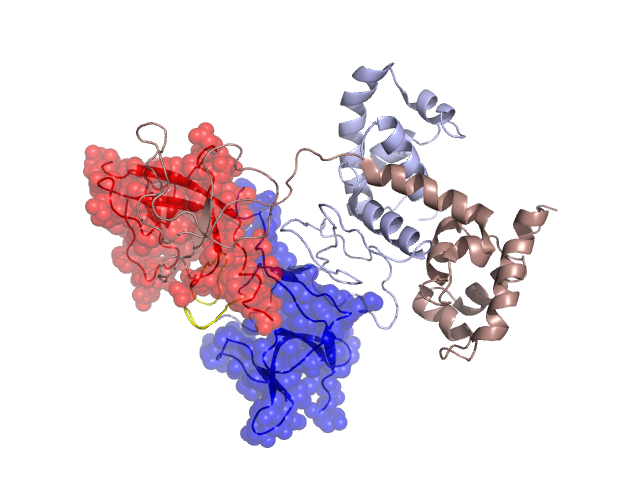
Structural Details of PDB entry 3BDN
Structural Details of PDB entry 3BDN
PDBid Chains Hinge Swapped Domain
3BDN
A,B
A:153-160,B:153-160
A:161-236,B:161-236
Swapped-domain interface residues and interactions:
Chains Residues
A
156 , 157 , 161 , 162 , 164 , 177 , 178 , 179 , 207 , 225 , 226 , 227 , 228 , 229 , 230 , 234 , 235 , 236 ,
B
155 , 161 , 162 , 164 , 177 , 178 , 179 , 224 , 225 , 226 , 227 , 228 , 229 , 230 , 232 , 234 , 235 ,
Non-swapped-domain interface residues and interactions:
Chains Residues
A
38 , 39 , 40 , 41 , 59 , 60 , 63 , 66 , 67 , 72 , 73 , 74 , 83 , 84 , 85 , 86 , 87 , 88 , 89 , 90 , 91 , 93 , 96 , 98 , 99 , 100 , 130 , 131 , 132 , 133 , 148 , 151 , 152 , 154 , 155 , 158 , 159 , 160 ,
B
60 , 63 , 67 , 70 , 71 , 72 , 73 , 74 , 75 , 81 , 83 , 84 , 85 , 86 , 87 , 88 , 89 , 90 , 91 , 96 , 98 , 99 , 100 , 115 , 116 , 117 , 120 , 129 , 131 , 132 , 133 , 148 , 151 , 152 , 154 , 156 , 157 , 158 , 159 , 160 ,
Mutations in critical regions:
Chains
Hinge
Domain swapped interface Non-swapped interface Swapped Domain
A No mutation No mutation No mutation GLY(197)ASP, B No mutation No mutation No mutation GLY(197)ASP,
HIDE output:
JMOL Visualization:
2D-plot:
JOY Structural annotation for hinge hinge and swapped domain:
JOY output: