Pfam Domains mapped on to the structure: 3BCO
No.
Chain ID
Pfam ID
Pfam Description
Linkout - Pfam
Linkout - CDD
1
A
PF00074
Pancreatic ribonuclease
PF00074
PF00074
Conserved Domain Database Superfamily Annotations: 3BCO
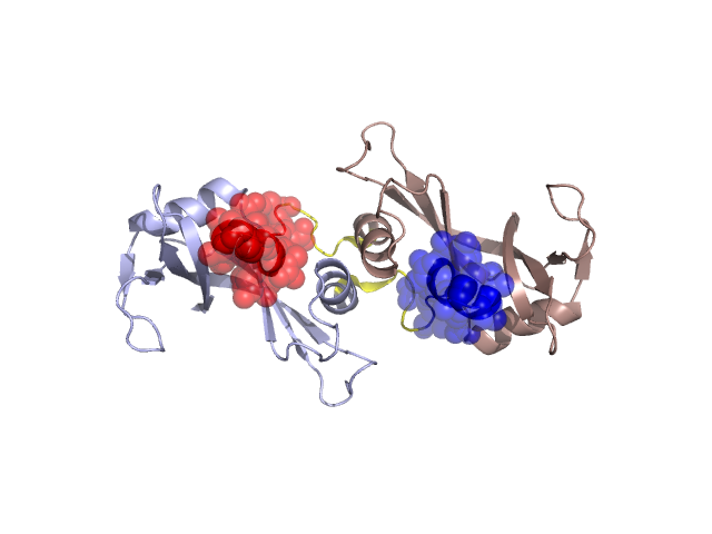
Structural Details of PDB entry 3BCO
Structural Details of PDB entry 3BCO
PDBid Chains Hinge Swapped Domain
3BCO
A,B
A:16-22,B:16-22
A:1-15,B:1-15
Swapped-domain interface residues and interactions:
Chains Residues
A
4 , 5 , 8 , 9 , 10 , 11 , 12 , 13 , 14 , 15 , 33 , 45 , 46 , 47 , 48 , 49 , 50 , 116 , 117 , 118 ,
B
4 , 5 , 8 , 9 , 10 , 11 , 12 , 13 , 14 , 15 , 33 , 45 , 46 , 47 , 48 , 49 , 50 , 51 , 116 , 117 , 118 ,
Non-swapped-domain interface residues and interactions:
Chains Residues
A
16 , 17 , 19 , 20 , 22 , 25 , 28 , 29 , 31 , 32 , 34 , 35 , 37 , 41 , 44 , 51 , 54 , 80 , 101 , 120 , 124 ,
B
18 , 19 , 22 , 25 , 28 , 29 , 31 , 32 , 34 , 35 , 41 , 44 , 54 , 80 , 101 , 120 ,
Mutations in critical regions:
Chains
Hinge
Domain swapped interface Non-swapped interface Swapped Domain
A ALA(19)PRO, No mutation ALA(19)PRO, GLN(28)LEU, No mutation B ALA(19)PRO, No mutation ALA(19)PRO, GLN(28)LEU, No mutation
HIDE output:
JMOL Visualization:
2D-plot:
JOY Structural annotation for hinge hinge and swapped domain:
JOY output: