Pfam Domains mapped on to the structure: 3B5K
No.
Chain ID
Pfam ID
Pfam Description
Linkout - Pfam
Linkout - CDD
1
A
PF02025
Interleukin 5
PF02025
PF02025
Gene Ontology Annotations: 3B5K
Conserved Domain Database Superfamily Annotations: 3B5K
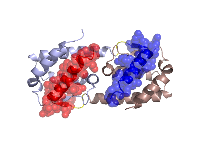
Structural Details of PDB entry 3B5K
Structural Details of PDB entry 3B5K
PDBid Chains Hinge Swapped Domain
3B5K
A,B
A:87-89,B:87-89
A:90-111,B:90-110
Swapped-domain interface residues and interactions:
Chains Residues
A
8 , 11 , 15 , 26 , 31 , 32 , 33 , 34 , 35 , 41 , 90 , 91 , 92 , 93 , 94 , 96 , 97 , 98 , 99 , 100 , 101 , 102 , 103 , 104 , 105 , 106 , 107 , 108 , 110 , 111 ,
B
8 , 11 , 15 , 26 , 29 , 31 , 32 , 33 , 34 , 35 , 41 , 90 , 91 , 92 , 93 , 94 , 95 , 96 , 97 , 98 , 99 , 100 , 101 , 102 , 103 , 104 , 105 , 106 , 107 , 108 ,
Non-swapped-domain interface residues and interactions:
Chains Residues
A
18 , 22 , 25 , 28 , 29 , 36 , 37 , 38 , 39 , 40 , 42 , 43 , 44 , 45 , 47 , 48 , 52 , 55 , 72 , 75 , 78 , 79 , 82 , 83 , 85 , 86 , 87 , 89 ,
B
7 , 14 , 18 , 22 , 25 , 28 , 36 , 37 , 38 , 39 , 40 , 42 , 43 , 44 , 45 , 46 , 47 , 48 , 51 , 52 , 54 , 55 , 72 , 75 , 78 , 79 , 82 , 83 , 85 , 86 , 87 , 89 ,
Mutations in critical regions:
Chains
Hinge
Domain swapped interface Non-swapped interface Swapped Domain
A No mutation LEU(31)MET, LEU(107)MET, No mutation LEU(107)MET, B No mutation LEU(31)MET, LEU(107)MET, THR(7)MET, LEU(107)MET,
HIDE output:
JMOL Visualization:
2D-plot:
JOY Structural annotation for hinge hinge and swapped domain:
JOY output: