Home BrowseSearch Help Group

Pfam Domains mapped on to the structure: 2YXH

No. Chain ID Pfam ID Pfam Description Linkout - Pfam Linkout - CDD
1 A PF03819 MazG nucleotide pyrophosphohydrolase domain PF03819 PF03819

Conserved Domain Database Superfamily Annotations: 2YXH

No. PDB ID PSSM ID CDD Accession Superfamily Short Name Linkout - CDD
1 2YXH 212135 cd11528 NTP-PPase_MazG_Nterm - cl16941
2 2YXH 212390 cl16941 NTP-PPase superfamily - -
Structural Details of PDB entry 2YXH

Structural Details of PDB entry 2YXH

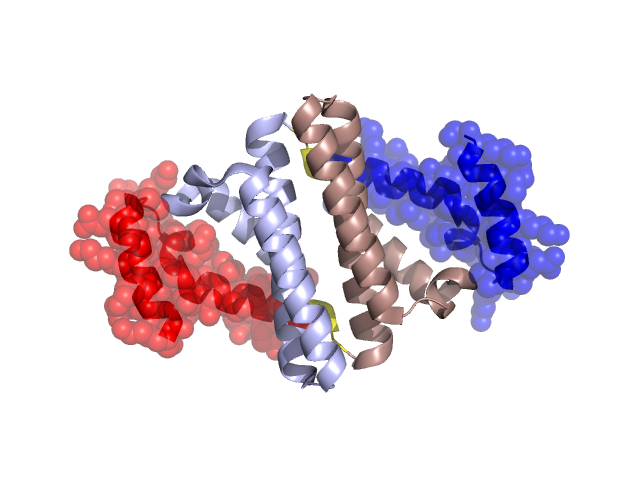
PDBid Chains Hinge Swapped Domain
2YXH A,B A:74-77,B:74-77 A:78-114,B:78-115

Swapped-domain interface residues and interactions:

Chains Residues
A 5, 11, 12, 15, 16, 17, 18, 51, 52, 54, 55, 56, 58, 78, 79, 80, 81, 82, 83, 84, 86, 87, 88, 91, 93, 94, 95, 97, 99, 100, 102, 105, 114,
B 2, 5, 11, 12, 15, 16, 17, 18, 51, 52, 54, 55, 56, 78, 79, 80, 82, 83, 84, 86, 87, 91, 93, 94, 95, 97, 99, 100, 102, 105,

Non-swapped-domain interface residues and interactions:

Chains Residues
A 2, 4, 6, 8, 21, 24, 25, 28, 31, 32, 35, 38, 39, 42, 45, 47, 50, 59, 61, 62, 64, 65, 68, 72, 74, 76, 77,
B 4, 6, 8, 14, 21, 24, 25, 28, 31, 32, 35, 38, 39, 42, 45, 47, 50, 58, 59, 61, 62, 64, 65, 68, 72, 74, 76, 77,

Swapped domains are represented using trasperent spheres.
Non-swapped part is represented using light color and cartoon representation.
Hinge region is shown in yellow color.

Mutations in critical regions:

Chains Hinge Domain swapped interface Non-swapped interface Swapped Domain
ANo mutationNo mutationNo mutationNo mutation
BNo mutationNo mutationNo mutationNo mutation

HIDE output:

Homologues found through HIDE algorithm

JMOL Visualization:

Jmol_logo            Jmol_logo            Jmol_logo

2D-plot:

A:2YXH B:2YXH

JOY Structural annotation for hinge hinge and swapped domain:

Hinge Swapped domain

JOY output:

ali file:2YXH.ali
atm file:2YXH.atm
cof file:2YXH.cof
hbd file:2YXH.hbd
html file:2YXH.html
pdb file:2YXH.pdb
ps file:2YXH.ps
psa file:2YXH.psa
rtf file:2YXH.rtf
sst file:2YXH.sst
tem file:2YXH.tem