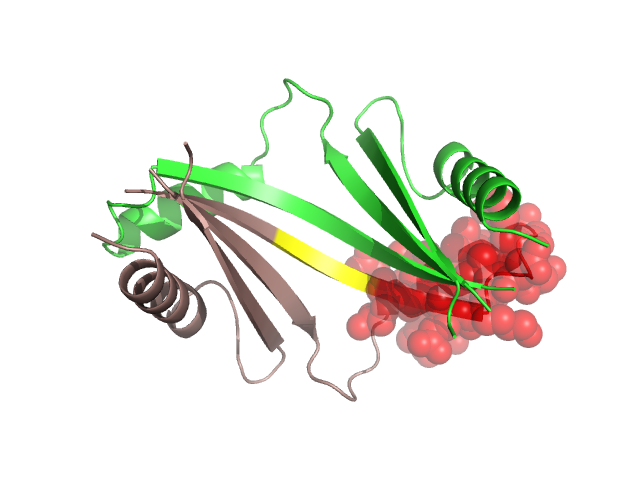
Structural Details of PDB entry 2YX5
Structural Details of PDB entry 2YX5
PDBid Chains Hinge Swapped Domain
2YX5
A,B
A:12-17,B:12-17;A:39-42,B:39-42
A:18-38,B:18-38
Swapped-domain interface residues and interactions:
Chains Residues
A
18 , 22 , 25 , 26 , 29 , 31 , 33 , 34 , 35 , 36 , 37 , 38 ,
D
43 , 44 , 45 , 46 , 47 , 48 ,
Non-swapped-domain interface residues and interactions:
Chains Residues
A
1 , 7 , 39 , 40 , 41 , 42 , 43 , 44 , 45 , 46 , 47 , 48 , 58 , 61 , 62 , 65 , 66 , 67 , 83 ,
D
1 , 7 , 18 , 22 , 25 , 26 , 29 , 31 , 33 , 34 , 35 , 36 , 37 , 38 , 39 , 40 , 41 , 42 , 58 , 61 , 62 , 65 , 66 , 67 ,