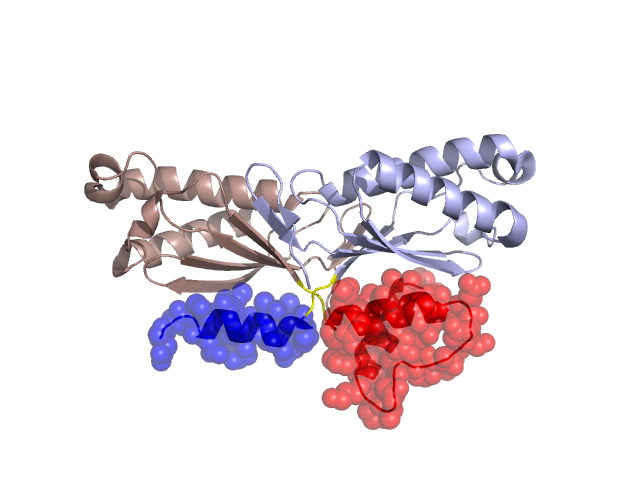
Structural Details of PDB entry 2VIH
Structural Details of PDB entry 2VIH
PDBid Chains Hinge Swapped Domain
2VIH
A,B
A:116-118,B:116-118
A:119-155,B:119-155
Swapped-domain interface residues and interactions:
Chains Residues
A
57 , 59 , 60 , 61 , 66 , 116 , 117 , 118 , 119 , 120 , 121 , 124 , 125 , 127 , 128 , 129 , 131 , 132 , 146 , 150 , 153 , 154 , 155 ,
B
57 , 59 , 60 , 61 , 66 , 116 , 117 , 118 , 119 , 120 , 121 , 124 , 125 , 127 , 128 , 129 , 131 , 132 ,
Non-swapped-domain interface residues and interactions:
Chains Residues
A
7 , 12 , 13 , 14 , 16 , 17 , 18 , 19 , 20 , 56 , 68 , 73 , 74 , 77 , 78 , 109 , 110 , 111 , 112 , 113 , 114 , 115 ,
B
5 , 7 , 12 , 13 , 14 , 16 , 17 , 18 , 19 , 20 , 22 , 28 , 37 , 56 , 64 , 68 , 73 , 74 , 77 , 78 , 109 , 110 , 111 , 112 , 113 , 114 , 115 ,