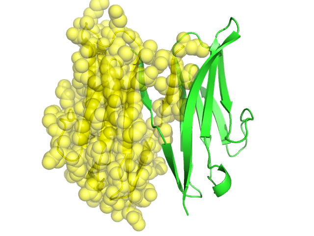
Pfam Domains mapped on to the structure: 2VAJ No. Chain ID Pfam ID Pfam Description Linkout - Pfam Linkout - CDD 1 A PF13895 Immunoglobulin domain PF13895 PF13895 2 A PF00047 Immunoglobulin domain PF00047 PF00047 Conserved Domain Database Superfamily Annotations: 2VAJ No. PDB ID PSSM ID CDD Accession Superfamily Short Name Linkout - CDD 1 2VAJ 212623 cl11960 Ig superfamily - - 2 2VAJ 212623 cl11960 Ig superfamily - - Structural Details of PDB entry 2VAJ Structural Details of PDB entry 2VAJ PDBid Chains Hinge Swapped Domain 2VAJ A,B A:-,B:- A:1--1,B:1--1 Swapped-domain interface residues and interactions: Chains Residues Non-swapped-domain interface residues and interactions: Chains Residues A 1, 2, 3, 4, 5, 6, 7, 8, 9, 10, 11, 16, 18, 19, 20, 21, 22, 23, 24, 25, 26, 75, 77, 83, 84, 85, 87, 92, 93, B 1, 2, 3, 4, 5, 6, 7, 8, 9, 10, 11, 16, 18, 19, 20, 21, 22, 23, 24, 25, 26, 75, 77, 83, 84, 85, 87, 92, Swapped domains are represented using trasperent spheres. Non-swapped part is represented using light color and cartoon representation. Hinge region is shown in yellow color. Mutations in critical regions: Chains Hinge Domain swapped interface Non-swapped interface Swapped Domain HIDE output: Homologues found through HIDE algorithm JMOL Visualization: 2D-plot: A:2VAJ B:2VAJ JOY Structural annotation for hinge hinge and swapped domain: Hinge Swapped domain JOY output: ali file:2VAJ.ali atm file:2VAJ.atm cof file:2VAJ.cof hbd file:2VAJ.hbd html file:2VAJ.html pdb file:2VAJ.pdb ps file:2VAJ.ps psa file:2VAJ.psa rtf file:2VAJ.rtf sst file:2VAJ.sst tem file:2VAJ.tem