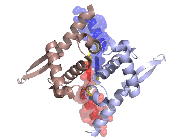
Structural Details of PDB entry 2V79
Structural Details of PDB entry 2V79
PDBid Chains Hinge Swapped Domain
2V79
A,B
A:17-20,B:17-20
A:1-16,B:1-16
Swapped-domain interface residues and interactions:
Chains Residues
A
1 , 2 , 5 , 9 , 12 , 13 , 14 , 15 , 16 , 17 , 18 , 19 , 20 , 111 ,
B
1 , 5 , 9 , 12 , 13 , 14 , 15 , 16 , 18 , 19 , 20 , 111 ,
Non-swapped-domain interface residues and interactions:
Chains Residues
A
22 , 23 , 25 , 26 , 31 , 32 , 33 , 35 , 36 , 39 , 40 , 43 , 60 , 61 , 62 , 107 , 110 , 114 , 115 ,
B
17 , 22 , 23 , 25 , 26 , 27 , 31 , 32 , 33 , 36 , 39 , 40 , 43 , 60 , 61 , 62 , 107 , 110 , 114 ,