Pfam Domains mapped on to the structure: 2TOH
No.
Chain ID
Pfam ID
Pfam Description
Linkout - Pfam
Linkout - CDD
1
A
PF00351
Biopterin-dependent aromatic amino acid hydroxylase
PF00351
PF00351
Conserved Domain Database Superfamily Annotations: 2TOH
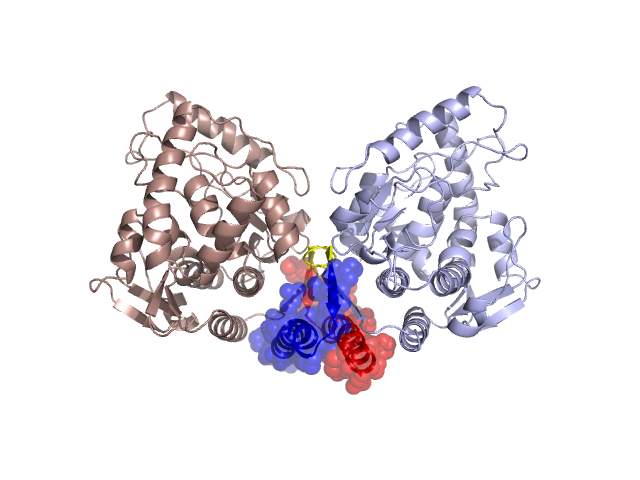
Structural Details of PDB entry 2TOH
Structural Details of PDB entry 2TOH
PDBid Chains Hinge Swapped Domain
2TOH
A,B
A:462-466,B:462-466
A:467-498,B:467-498
Swapped-domain interface residues and interactions:
Chains Residues
A
450 , 454 , 455 , 456 , 457 , 459 , 470 , 471 , 473 , 476 , 478 , 479 , 480 , 481 , 482 , 483 , 485 , 486 , 487 , 489 , 490 , 492 , 493 , 494 , 496 , 497 , 498 ,
B
450 , 454 , 455 , 456 , 457 , 459 , 470 , 471 , 473 , 476 , 478 , 479 , 480 , 481 , 482 , 483 , 485 , 486 , 487 , 489 , 490 , 492 , 493 , 494 , 496 , 497 ,
Non-swapped-domain interface residues and interactions:
Chains Residues
A
170 , 172 , 219 , 281 , 282 , 284 , 285 , 307 , 343 , 347 , 439 , 442 , 446 , 458 , 460 , 462 , 463 , 464 ,
B
170 , 172 , 219 , 281 , 282 , 284 , 285 , 307 , 343 , 347 , 439 , 442 , 446 , 458 , 460 , 462 , 463 , 464 ,
Mutations in critical regions:
Chains
Hinge
Domain swapped interface Non-swapped interface Swapped Domain
A No mutation No mutation No mutation No mutation B No mutation No mutation No mutation No mutation
HIDE output:
JMOL Visualization:
2D-plot:
JOY Structural annotation for hinge hinge and swapped domain:
JOY output: