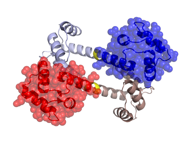
Pfam Domains mapped on to the structure: 2R28 No. Chain ID Pfam ID Pfam Description Linkout - Pfam Linkout - CDD 1 A PF00036 EF hand PF00036 PF00036 2 A PF00036 EF hand PF00036 PF00036 3 A PF00036 EF hand PF00036 PF00036 4 A PF00036 EF hand PF00036 PF00036 5 A PF13499 EF-hand domain pair PF13499 PF13499 6 A PF13499 EF-hand domain pair PF13499 PF13499 7 A PF13833 EF-hand domain pair PF13833 PF13833 8 A PF13833 EF-hand domain pair PF13833 PF13833 9 A PF13833 EF-hand domain pair PF13833 PF13833 10 A PF08726 Ca2+ insensitive EF hand PF08726 PF08726 11 A PF12763 Cytoskeletal-regulatory complex EF hand PF12763 PF12763 Structural Details of PDB entry 2R28 Structural Details of PDB entry 2R28 PDBid Chains Hinge Swapped Domain 2R28 A,B A:78-82,B:78-82 A:83-148,B:83-148 Swapped-domain interface residues and interactions: Chains Residues A 19, 39, 40, 84, 91, 92, 95, 109, 112, 113, 114, 148, B 19, 22, 39, 40, 41, 84, 91, 92, 95, 112, 113, 114, Non-swapped-domain interface residues and interactions: Chains Residues A 22, 35, 36, 41, B 35, Swapped domains are represented using trasperent spheres. Non-swapped part is represented using light color and cartoon representation. Hinge region is shown in yellow color. Mutations in critical regions: Chains Hinge Domain swapped interface Non-swapped interface Swapped Domain ANo mutationNo mutationNo mutationNo mutation BNo mutationNo mutationNo mutationNo mutation HIDE output: Homologues found through HIDE algorithm JMOL Visualization: 2D-plot: A:2R28 B:2R28 JOY Structural annotation for hinge hinge and swapped domain: Hinge Swapped domain JOY output: ali file:2R28.ali atm file:2R28.atm cof file:2R28.cof hbd file:2R28.hbd html file:2R28.html pdb file:2R28.pdb ps file:2R28.ps psa file:2R28.psa rtf file:2R28.rtf sst file:2R28.sst tem file:2R28.tem