Pfam Domains mapped on to the structure: 2QUD
No.
Chain ID
Pfam ID
Pfam Description
Linkout - Pfam
Linkout - CDD
1
A
PF09063
Phage PP7 coat protein
PF09063
PF09063
Conserved Domain Database Superfamily Annotations: 2QUD
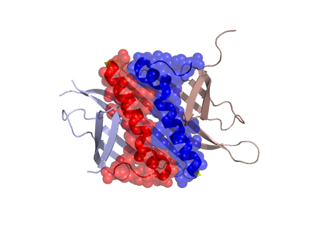
Structural Details of PDB entry 2QUD
Structural Details of PDB entry 2QUD
PDBid Chains Hinge Swapped Domain
2QUD
A,B
A:92-95,B:92-95
A:96-127,B:96-127
Swapped-domain interface residues and interactions:
Chains Residues
A
2 , 3 , 4 , 6 , 7 , 61 , 85 , 90 , 93 , 96 , 98 , 99 , 100 , 101 , 102 , 103 , 104 , 105 , 106 , 107 , 108 , 109 , 110 , 111 , 112 , 114 , 115 , 116 , 118 , 119 , 120 , 121 , 122 , 124 , 125 , 126 , 127 ,
B
2 , 3 , 4 , 6 , 8 , 44 , 61 , 85 , 90 , 96 , 98 , 99 , 100 , 101 , 102 , 103 , 104 , 105 , 106 , 107 , 108 , 109 , 110 , 111 , 112 , 114 , 115 , 116 , 118 , 119 , 120 , 121 , 122 , 124 , 125 , 126 ,
Non-swapped-domain interface residues and interactions:
Chains Residues
A
0 , 5 , 8 , 13 , 25 , 27 , 38 , 40 , 42 , 44 , 53 , 55 , 57 , 59 , 63 , 79 , 80 , 81 , 82 , 83 , 84 , 86 , 87 , 88 , 89 , 91 , 94 , 95 ,
B
5 , 7 , 13 , 25 , 27 , 38 , 40 , 42 , 53 , 55 , 57 , 59 , 63 , 79 , 80 , 81 , 82 , 83 , 84 , 86 , 87 , 88 , 89 , 91 , 93 , 94 , 95 ,
Mutations in critical regions:
Chains
Hinge
Domain swapped interface Non-swapped interface Swapped Domain
A No mutation No mutation No mutation No mutation B No mutation No mutation No mutation No mutation
HIDE output:
JMOL Visualization:
2D-plot:
JOY Structural annotation for hinge hinge and swapped domain:
JOY output: