Structural Details of PDB entry 2QSC
Structural Details of PDB entry 2QSC
PDBid Chains Hinge Swapped Domain
2QSC
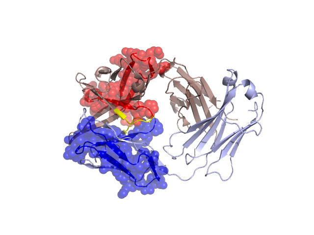
H,L
H:42-46,L:41-45
H:1-41,L:1-40
Swapped-domain interface residues and interactions:
Non-swapped-domain interface residues and interactions:
Chains Residues
H
44 , 45 , 47 , 50 , 58 , 91 , 98 , 100 , 101 , 103 , 104 , 105 , 122 , 123 , 124 , 125 , 126 , 127 , 137 , 139 , 143 , 145 , 172 , 173 , 174 , 175 , 177 , 178 , 179 , 188 , 190 , 192 , 221 ,
L
41 , 42 , 43 , 44 , 46 , 49 , 50 , 55 , 87 , 89 , 91 , 94 , 95 , 96 , 98 , 99 , 100 , 116 , 118 , 119 , 121 , 123 , 124 , 127 , 129 , 131 , 133 , 135 , 137 , 138 , 160 , 161 , 162 , 163 , 164 , 167 , 174 , 176 , 178 , 180 , 214 ,