Home BrowseSearch Help Group

Pfam Domains mapped on to the structure: 2QPV

No. Chain ID Pfam ID Pfam Description Linkout - Pfam Linkout - CDD
1 A PF10604 Polyketide cyclase / dehydrase and lipid transport PF10604 PF10604

Conserved Domain Database Superfamily Annotations: 2QPV

No. PDB ID PSSM ID CDD Accession Superfamily Short Name Linkout - CDD
1 2QPV 209878 cl14643 SRPBCC superfamily - -
Structural Details of PDB entry 2QPV

Structural Details of PDB entry 2QPV

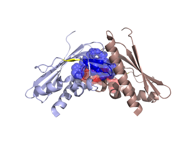
PDBid Chains Hinge Swapped Domain
2QPV A,B A:45-47,B:45-47;A:60-62,B:60-62 A:48-59,B:48-59

Swapped-domain interface residues and interactions:

Chains Residues
A 34, 35, 36, 37, 49, 51, 52, 53, 54, 55, 56, 57, 58, 59, 75, 115, 116, 119, 120, 123,
B 34, 35, 36, 37, 49, 51, 52, 53, 54, 55, 56, 57, 58, 59, 75, 112, 116, 119, 120, 123,

Non-swapped-domain interface residues and interactions:

Chains Residues
A 38, 39, 44, 77, 78, 79, 80, 81, 85, 105, 106, 111, 112, 118, 133,
B 38, 39, 44, 77, 78, 79, 81, 85, 105, 106, 107, 111, 115, 118,

Swapped domains are represented using trasperent spheres.
Non-swapped part is represented using light color and cartoon representation.
Hinge region is shown in yellow color.

Mutations in critical regions:

Chains Hinge Domain swapped interface Non-swapped interface Swapped Domain
ANo mutationNo mutationNo mutationNo mutation
BNo mutationNo mutationNo mutationNo mutation

HIDE output:

Homologues found through HIDE algorithm

JMOL Visualization:

Jmol_logo            Jmol_logo            Jmol_logo

2D-plot:

A:2QPV B:2QPV

JOY Structural annotation for hinge hinge and swapped domain:

Hinge Swapped domain

JOY output:

ali file:2QPV.ali
atm file:2QPV.atm
cof file:2QPV.cof
hbd file:2QPV.hbd
html file:2QPV.html
pdb file:2QPV.pdb
ps file:2QPV.ps
psa file:2QPV.psa
rtf file:2QPV.rtf
sst file:2QPV.sst
tem file:2QPV.tem