Pfam Domains mapped on to the structure: 2QNU
No.
Chain ID
Pfam ID
Pfam Description
Linkout - Pfam
Linkout - CDD
1
A
PF09867
Uncharacterized protein conserved in bacteria (DUF2094)
PF09867
PF09867
Conserved Domain Database Superfamily Annotations: 2QNU
Structural Details of PDB entry 2QNU
Structural Details of PDB entry 2QNU
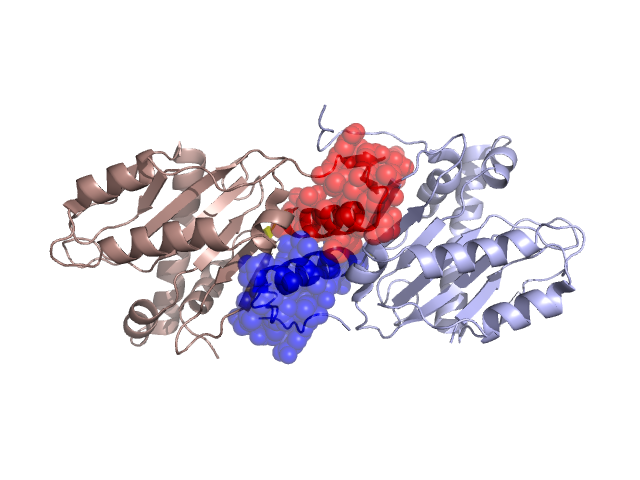
PDBid Chains Hinge Swapped Domain
2QNU
A,B
A:146-147,B:146-147;A:179-180,B:179-180
A:148-178,B:148-178
Swapped-domain interface residues and interactions:
Chains Residues
A
102 , 105 , 106 , 107 , 149 , 150 , 151 , 152 , 153 , 154 , 156 , 157 , 158 , 159 , 160 , 162 , 163 , 165 , 168 , 169 , 170 , 172 , 173 , 175 , 176 , 193 , 194 , 195 , 197 , 198 ,
B
102 , 105 , 106 , 107 , 153 , 156 , 157 , 158 , 159 , 160 , 162 , 163 , 165 , 168 , 169 , 172 , 173 , 175 , 176 , 193 , 194 , 195 , 197 , 198 , 215 , 216 , 217 , 218 , 219 , 220 , 221 , 222 , 223 ,
Non-swapped-domain interface residues and interactions:
Chains Residues
A
97 , 98 , 103 , 109 , 146 , 147 , 184 , 192 , 203 , 209 , 214 ,
B
103 , 109 , 184 , 192 , 203 , 209 , 213 , 224 ,
Mutations in critical regions:
Chains
Hinge
Domain swapped interface Non-swapped interface Swapped Domain
A No mutation No mutation No mutation No mutation B No mutation No mutation No mutation No mutation B No mutation No mutation No mutation No mutation
HIDE output:
JMOL Visualization:
2D-plot:
JOY Structural annotation for hinge hinge and swapped domain:
JOY output: