Pfam Domains mapped on to the structure: 2Q8G
No.
Chain ID
Pfam ID
Pfam Description
Linkout - Pfam
Linkout - CDD
1
A
PF10436
Mitochondrial branched-chain alpha-ketoacid dehydrogenase kinase
PF10436
PF10436
2
A
PF02518
Histidine kinase-, DNA gyrase B-, and HSP90-like ATPase
PF02518
PF02518
Conserved Domain Database Superfamily Annotations: 2Q8G
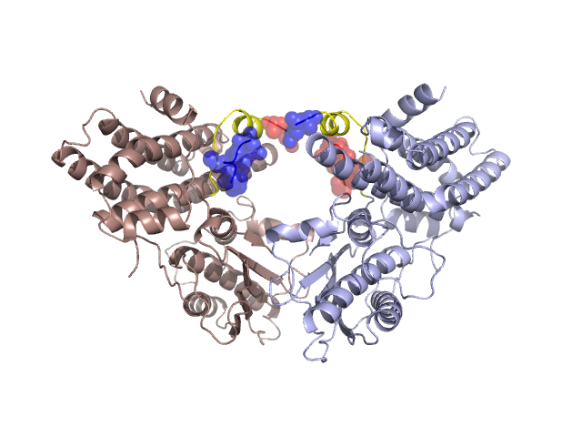
Structural Details of PDB entry 2Q8G
Structural Details of PDB entry 2Q8G
PDBid Chains Hinge Swapped Domain
2Q8G
A,B
A:399-412,B:399-412
A:413-423,B:413-423
Swapped-domain interface residues and interactions:
Chains Residues
A
399 , 400 , 401 , 402 , 404 , 406 , 418 , 419 , 420 , 421 , 423 ,
B
397 , 399 , 400 , 401 , 402 , 404 , 406 , 418 , 419 , 420 , 421 ,
Non-swapped-domain interface residues and interactions:
Chains Residues
A
53 , 56 , 179 , 183 , 186 , 253 , 255 , 257 , 304 , 306 , 308 , 309 , 310 , 311 , 313 , 315 , 324 , 325 , 326 , 370 , 372 , 374 , 376 , 377 , 378 , 379 , 380 , 381 , 384 , 386 , 388 , 390 , 397 ,
B
53 , 56 , 179 , 183 , 186 , 253 , 255 , 257 , 304 , 306 , 308 , 309 , 310 , 311 , 313 , 315 , 324 , 325 , 326 , 370 , 372 , 374 , 376 , 377 , 378 , 379 , 380 , 381 , 384 , 386 , 388 , 390 ,
Mutations in critical regions:
Chains
Hinge
Domain swapped interface Non-swapped interface Swapped Domain
A No mutation No mutation No mutation No mutation B No mutation No mutation No mutation No mutation
HIDE output:
JMOL Visualization:
2D-plot:
JOY Structural annotation for hinge hinge and swapped domain:
JOY output: