Pfam Domains mapped on to the structure: 2Q4P
No.
Chain ID
Pfam ID
Pfam Description
Linkout - Pfam
Linkout - CDD
1
A
PF03819
MazG nucleotide pyrophosphohydrolase domain
PF03819
PF03819
Conserved Domain Database Superfamily Annotations: 2Q4P
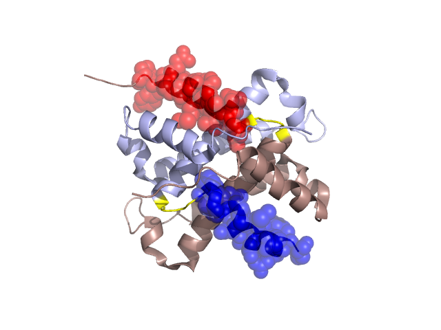
Structural Details of PDB entry 2Q4P
Structural Details of PDB entry 2Q4P
PDBid Chains Hinge Swapped Domain
2Q4P
A,B
A:110-113,B:110-113
A:114-130,B:114-130
Swapped-domain interface residues and interactions:
Chains Residues
A
25 , 26 , 27 , 28 , 29 , 94 , 97 , 101 , 114 , 115 , 116 , 117 , 118 , 119 , 121 , 125 , 126 , 129 ,
B
25 , 26 , 29 , 41 , 93 , 94 , 96 , 97 , 98 , 114 , 115 , 116 , 117 , 118 , 119 , 121 , 124 , 125 , 126 , 128 , 129 ,
Non-swapped-domain interface residues and interactions:
Chains Residues
A
23 , 24 , 30 , 31 , 32 , 34 , 35 , 36 , 37 , 38 , 41 , 45 , 49 , 50 , 52 , 53 , 54 , 57 , 60 , 61 , 63 , 64 , 67 , 68 , 71 , 72 , 74 , 76 , 80 , 81 , 82 , 89 , 92 , 93 , 96 , 98 , 99 , 100 , 102 , 103 , 106 , 107 , 108 , 109 , 110 , 111 , 112 , 113 , 131 , 132 , 134 ,
B
23 , 24 , 27 , 30 , 31 , 32 , 34 , 35 , 37 , 44 , 45 , 46 , 52 , 53 , 54 , 57 , 60 , 61 , 64 , 67 , 68 , 71 , 72 , 74 , 76 , 80 , 81 , 82 , 89 , 92 , 99 , 100 , 101 , 103 , 106 , 107 , 108 , 109 , 110 , 111 , 112 , 113 ,
Mutations in critical regions:
Chains
Hinge
Domain swapped interface Non-swapped interface Swapped Domain
A No mutation No mutation No mutation No mutation B No mutation No mutation No mutation No mutation
HIDE output:
JMOL Visualization:
2D-plot:
JOY Structural annotation for hinge hinge and swapped domain:
JOY output: