Home BrowseSearch Help Group

Pfam Domains mapped on to the structure: 2PVA

No. Chain ID Pfam ID Pfam Description Linkout - Pfam Linkout - CDD
1 A PF02275 Linear amide C-N hydrolases, choloylglycine hydrolase family PF02275 PF02275

Conserved Domain Database Superfamily Annotations: 2PVA

No. PDB ID PSSM ID CDD Accession Superfamily Short Name Linkout - CDD
1 2PVA 48435 cd00542 Ntn_PVA - cl00467
2 2PVA 212223 cl00467 Ntn_hydrolase superfamily - -
3 2PVA 212223 cl00467 Ntn_hydrolase superfamily - -
Structural Details of PDB entry 2PVA

Structural Details of PDB entry 2PVA

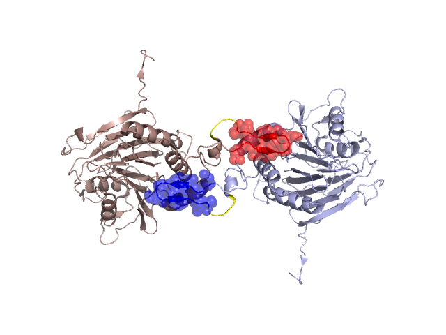
PDBid Chains Hinge Swapped Domain
2PVA A,B A:193-197,B:193-197;A:210-213,B:210-213 A:198-209,B:198-209

Swapped-domain interface residues and interactions:

Chains Residues
A 170, 187, 188, 190, 200, 201, 202, 204, 205, 207, 208, 209, 237,
B 170, 187, 188, 190, 200, 201, 202, 205, 207, 208, 209, 237,

Non-swapped-domain interface residues and interactions:

Chains Residues
A 183, 184, 186, 189, 191, 197, 210, 217, 218, 233, 234, 238, 332,
B 168, 183, 184, 186, 189, 191, 197, 210, 214, 217, 218, 233, 234, 238,

Swapped domains are represented using trasperent spheres.
Non-swapped part is represented using light color and cartoon representation.
Hinge region is shown in yellow color.

Mutations in critical regions:

Chains Hinge Domain swapped interface Non-swapped interface Swapped Domain
ANo mutationNo mutationNo mutationNo mutation
BNo mutationNo mutationNo mutationNo mutation

HIDE output:

Homologues found through HIDE algorithm

JMOL Visualization:

Jmol_logo            Jmol_logo            Jmol_logo

2D-plot:

A:2PVA B:2PVA

JOY Structural annotation for hinge hinge and swapped domain:

Hinge Swapped domain

JOY output:

ali file:2PVA.ali
atm file:2PVA.atm
cof file:2PVA.cof
hbd file:2PVA.hbd
html file:2PVA.html
pdb file:2PVA.pdb
ps file:2PVA.ps
psa file:2PVA.psa
rtf file:2PVA.rtf
sst file:2PVA.sst
tem file:2PVA.tem