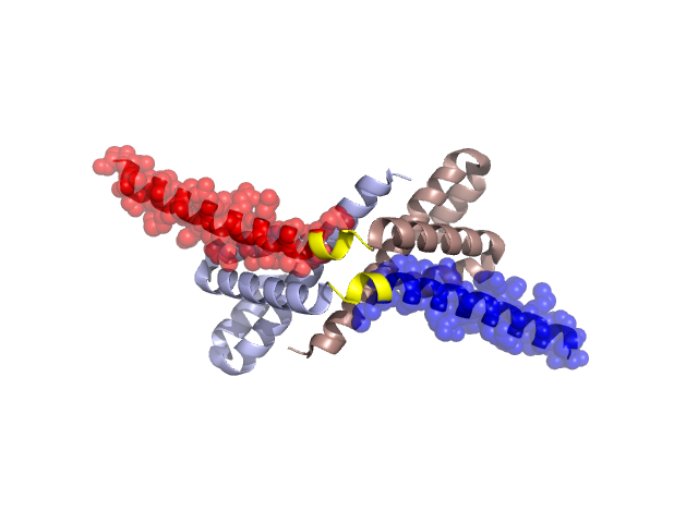
Structural Details of PDB entry 2PEO
Structural Details of PDB entry 2PEO
PDBid Chains Hinge Swapped Domain
2PEO
A,B
A:83-87,B:83-87
A:88-115,B:88-114
Swapped-domain interface residues and interactions:
Chains Residues
A
11 , 15 , 71 , 75 , 78 , 79 , 88 , 89 , 90 , 92 , 93 , 94 , 96 , 97 , 100 , 101 , 103 , 104 , 107 , 115 ,
B
11 , 14 , 15 , 71 , 75 , 78 , 79 , 88 , 89 , 90 , 92 , 93 , 94 , 96 , 97 , 100 , 101 , 104 ,
Non-swapped-domain interface residues and interactions:
Chains Residues
A
1 , 3 , 6 , 7 , 9 , 10 , 13 , 14 , 17 , 18 , 21 , 25 , 29 , 53 , 57 , 60 , 64 , 68 , 73 , 74 , 76 , 77 , 80 , 81 , 82 , 85 , 86 ,
B
1 , 3 , 6 , 7 , 9 , 10 , 13 , 17 , 18 , 21 , 25 , 28 , 29 , 53 , 56 , 57 , 60 , 61 , 64 , 68 , 72 , 74 , 77 , 80 , 81 , 82 , 85 , 86 ,