Home BrowseSearch Help Group

Structural Details of PDB entry 2PD1

Structural Details of PDB entry 2PD1

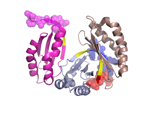
PDBid Chains Hinge Swapped Domain
2PD1 A,D,C,B A:90-92,D:90-92,C:90-92,B:90-92 A:93-100,D:93-100,C:93-100,B:93-100

Swapped-domain interface residues and interactions:

Chains Residues
A 21, 25, 28, 32, 38, 39, 40, 41, 42, 43, 44, 93, 94, 95, 96, 97, 98, 99, 100, 101,
B 100,
C 25, 28, 31, 32, 38, 39, 40, 41, 42, 43, 44, 92, 93, 94, 95, 96, 97, 98, 99,

Non-swapped-domain interface residues and interactions:

Chains Residues
A 7, 9, 31, 37, 45, 47, 53, 55, 89, 90, 91, 92,
B 66, 67, 71, 72, 76, 79, 83, 84, 85, 86, 87, 88, 89, 90, 92,
C 7, 9, 21, 37, 45, 53, 63, 66, 67, 71, 76, 79, 85, 87, 88, 89, 90, 91,

Swapped domains are represented using trasperent spheres.
Non-swapped part is represented using light color and cartoon representation.
Hinge region is shown in yellow color.

Mutations in critical regions:

Chains Hinge Domain swapped interface Non-swapped interface Swapped Domain
ANo mutationGLY(101)-, No mutationNo mutation
DNo mutationNo mutationNo mutationNo mutation
CNo mutationNo mutationNo mutationNo mutation
BNo mutationNo mutationNo mutationNo mutation

HIDE output:

Homologues found through HIDE algorithm

JMOL Visualization:

Jmol_logo            Jmol_logo            Jmol_logo

2D-plot:

A:2PD1 B:2PD1 C:2PD1 D:2PD1

JOY Structural annotation for hinge hinge and swapped domain:

Hinge Swapped domain

JOY output:

ali file:2PD1.ali
atm file:2PD1.atm
cof file:2PD1.cof
hbd file:2PD1.hbd
html file:2PD1.html
pdb file:2PD1.pdb
ps file:2PD1.ps
psa file:2PD1.psa
rtf file:2PD1.rtf
sst file:2PD1.sst
tem file:2PD1.tem