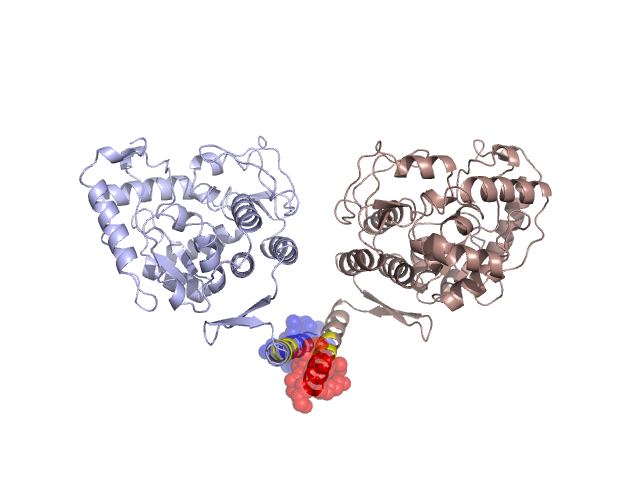
Pfam Domains mapped on to the structure: 2PAH No. Chain ID Pfam ID Pfam Description Linkout - Pfam Linkout - CDD 1 A PF00351 Biopterin-dependent aromatic amino acid hydroxylase PF00351 PF00351 Conserved Domain Database Superfamily Annotations: 2PAH No. PDB ID PSSM ID CDD Accession Superfamily Short Name Linkout - CDD 1 2PAH 48243 cd03347 eu_PheOH - cl01244 2 2PAH 207351 cl01244 arom_aa_hydroxylase superfamily - - Structural Details of PDB entry 2PAH Structural Details of PDB entry 2PAH PDBid Chains Hinge Swapped Domain 2PAH A,B A:437-441,B:437-441 A:442-452,B:442-452 Swapped-domain interface residues and interactions: Chains Residues A 431, 434, 435, 438, 442, 445, 448, 449, 451, 452, B 427, 431, 434, 435, 438, 442, 445, 448, 449, 451, Non-swapped-domain interface residues and interactions: Chains Residues A 366, 368, 370, 371, 427, 430, 437, 441, B 366, 368, 390, 430, 437, 441, Swapped domains are represented using trasperent spheres. Non-swapped part is represented using light color and cartoon representation. Hinge region is shown in yellow color. Mutations in critical regions: Chains Hinge Domain swapped interface Non-swapped interface Swapped Domain ANo mutationNo mutationNo mutationNo mutation BNo mutationNo mutationNo mutationNo mutation HIDE output: Homologues found through HIDE algorithm JMOL Visualization: 2D-plot: A:2PAH B:2PAH JOY Structural annotation for hinge hinge and swapped domain: Hinge Swapped domain JOY output: ali file:2PAH.ali atm file:2PAH.atm cof file:2PAH.cof hbd file:2PAH.hbd html file:2PAH.html pdb file:2PAH.pdb ps file:2PAH.ps psa file:2PAH.psa rtf file:2PAH.rtf sst file:2PAH.sst tem file:2PAH.tem