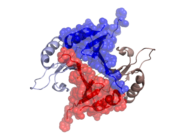
Structural Details of PDB entry 2P7O
Structural Details of PDB entry 2P7O
PDBid Chains Hinge Swapped Domain
2P7O
A,B
A:60-67,B:60-67
A:1-59,B:1-59
Swapped-domain interface residues and interactions:
Chains Residues
A
1 , 2 , 3 , 4 , 5 , 6 , 7 , 8 , 9 , 10 , 11 , 26 , 27 , 40 , 42 , 44 , 46 , 50 , 52 , 53 , 57 , 67 , 68 , 69 , 70 , 71 , 72 , 73 , 74 , 120 ,
B
1 , 2 , 3 , 4 , 5 , 6 , 7 , 8 , 9 , 10 , 11 , 26 , 44 , 46 , 50 , 52 , 53 , 55 , 57 , 67 , 68 , 69 , 70 , 71 , 72 , 73 , 74 , 75 ,
Non-swapped-domain interface residues and interactions:
Chains Residues
A
63 , 66 , 75 , 78 , 82 , 124 , 125 , 126 , 128 , 132 ,
B
63 , 66 , 82 , 115 , 124 , 125 , 126 , 128 , 129 ,