Pfam Domains mapped on to the structure: 2P10
No.
Chain ID
Pfam ID
Pfam Description
Linkout - Pfam
Linkout - CDD
1
A
PF03060
Nitronate monooxygenase
PF03060
PF03060
Gene Ontology Annotations: 2P10
Conserved Domain Database Superfamily Annotations: 2P10
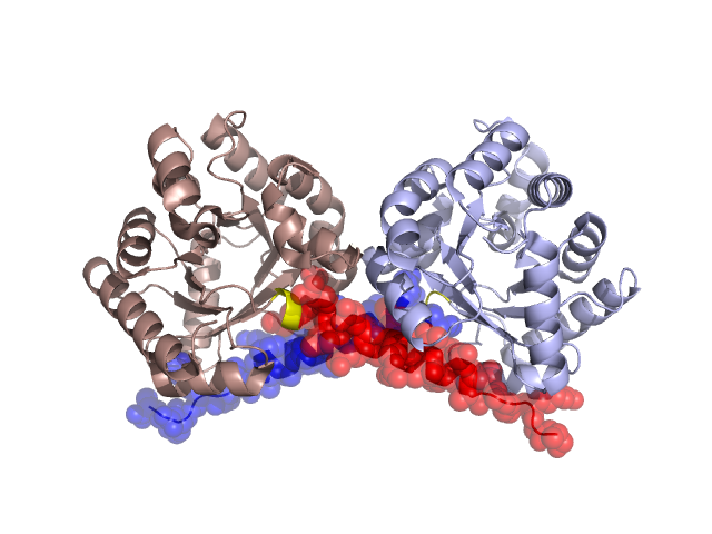
Structural Details of PDB entry 2P10
Structural Details of PDB entry 2P10
PDBid Chains Hinge Swapped Domain
2P10
A,B
A:258-260,B:258-260
A:261-284,B:261-285
Swapped-domain interface residues and interactions:
Chains Residues
A
27 , 28 , 30 , 38 , 39 , 42 , 43 , 45 , 46 , 47 , 48 , 239 , 243 , 246 , 247 , 262 , 263 , 265 , 266 , 267 , 269 , 270 , 271 , 273 , 274 , 275 , 276 , 277 , 278 , 280 , 281 , 282 , 283 , 284 ,
B
27 , 28 , 30 , 38 , 39 , 42 , 43 , 45 , 46 , 48 , 239 , 246 , 247 , 262 , 265 , 266 , 267 , 269 , 270 , 271 , 273 , 274 , 275 , 276 , 277 , 278 , 280 , 281 , 282 , 283 ,
Non-swapped-domain interface residues and interactions:
Chains Residues
A
26 , 36 , 37 , 41 , 60 , 62 , 63 , 64 , 85 , 86 , 89 , 90 , 242 , 250 , 252 , 255 , 260 ,
B
22 , 26 , 36 , 37 , 41 , 47 , 60 , 62 , 63 , 64 , 73 , 85 , 86 , 89 , 90 , 242 , 243 , 250 , 255 , 260 ,
Mutations in critical regions:
Chains
Hinge
Domain swapped interface Non-swapped interface Swapped Domain
A No mutation No mutation No mutation No mutation B No mutation No mutation No mutation No mutation
HIDE output:
JMOL Visualization:
2D-plot:
JOY Structural annotation for hinge hinge and swapped domain:
JOY output: