Home BrowseSearch Help Group

Pfam Domains mapped on to the structure: 2P06

No. Chain ID Pfam ID Pfam Description Linkout - Pfam Linkout - CDD
1 A PF03819 MazG nucleotide pyrophosphohydrolase domain PF03819 PF03819

Conserved Domain Database Superfamily Annotations: 2P06

No. PDB ID PSSM ID CDD Accession Superfamily Short Name Linkout - CDD
1 2P06 212140 cd11533 NTP-PPase_Af0060_like N cl16941
2 2P06 212390 cl16941 NTP-PPase superfamily N -
Structural Details of PDB entry 2P06

Structural Details of PDB entry 2P06

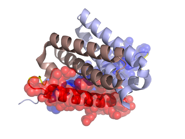
PDBid Chains Hinge Swapped Domain
2P06 A,B A:26-29,B:26-29 A:1-25,B:1-25

Swapped-domain interface residues and interactions:

Chains Residues
A 2, 3, 4, 6, 7, 9, 10, 11, 13, 15, 18, 79,
B 2, 3, 4, 6, 7, 9, 10, 11, 13, 15, 18, 79,

Non-swapped-domain interface residues and interactions:

Chains Residues
A 29, 30, 32, 41, 44, 45, 49, 51, 52, 55, 60, 63, 64, 67, 68, 70, 71, 72, 74, 76, 77, 78, 81, 82, 84,
B 1, 0, 30, 32, 41, 44, 45, 49, 51, 52, 55, 60, 63, 64, 67, 68, 70, 71, 72, 74, 76, 77, 78, 81, 82,

Swapped domains are represented using trasperent spheres.
Non-swapped part is reprented using light color and cartoon reprentation.
Hinge region is shown in yellow color.

Mutations in critical regions:

Chains Hinge Domain swapped interface Non-swapped interface Swapped Domain
ANo mutationNo mutationNo mutationNo mutation
BNo mutationNo mutationNo mutationNo mutation

HIDE output:

Homologues found through HIDE algorithm

JMOL Visualization:

Jmol_logo Jmol_logo

2D-plot:

3:2P06 A:2P06 B:2P06

JOY Structural annotation for hinge hinge and swapped domain:

Hinge Swapped domain

JOY output:

ali file:2P06.ali
atm file:2P06.atm
cof file:2P06.cof
hbd file:2P06.hbd
html file:2P06.html
pdb file:2P06.pdb
ps file:2P06.ps
psa file:2P06.psa
rtf file:2P06.rtf
sst file:2P06.sst
tem file:2P06.tem