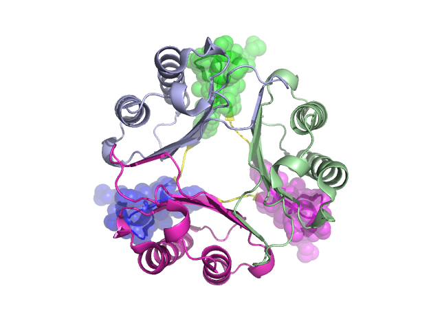
Pfam Domains mapped on to the structure: 2OS5
No.
Chain ID
Pfam ID
Pfam Description
Linkout - Pfam
Linkout - CDD
1
A
PF01187
Macrophage migration inhibitory factor (MIF)
PF01187
PF01187
Structural Details of PDB entry 2OS5
Structural Details of PDB entry 2OS5
PDBid Chains Hinge Swapped Domain
2OS5
A,D,C,B
A:100-102,D:100-102,C:100-102,B:100-102
A:103-118,D:103-118,C:103-118,B:103-118
Swapped-domain interface residues and interactions:
Chains Residues
B
69 , 72 , 73 , 76 , 77 , 80 , 91 , 92 , 94 , 95 , 96 , 97 , 98 , 104 , 105 , 106 , 107 , 108 , 109 , 110 , 111 , 112 , 115 , 118 ,
C
69 , 72 , 73 , 76 , 77 , 80 , 91 , 92 , 94 , 95 , 96 , 97 , 98 , 104 , 105 , 106 , 107 , 108 , 109 , 110 , 111 , 112 , 115 , 118 ,
D
69 , 72 , 73 , 76 , 77 , 80 , 91 , 92 , 94 , 95 , 96 , 97 , 98 , 104 , 105 , 106 , 107 , 108 , 109 , 110 , 111 , 112 , 115 ,
Non-swapped-domain interface residues and interactions:
Chains Residues
B
2 , 4 , 6 , 11 , 14 , 16 , 19 , 20 , 23 , 34 , 35 , 37 , 38 , 39 , 40 , 41 , 42 , 45 , 46 , 47 , 48 , 49 , 50 , 51 , 53 , 58 , 60 , 62 , 67 , 99 , 100 , 101 ,
C
2 , 4 , 11 , 14 , 16 , 19 , 20 , 23 , 35 , 37 , 38 , 39 , 40 , 41 , 42 , 45 , 46 , 47 , 48 , 49 , 50 , 51 , 53 , 58 , 60 , 62 , 67 , 99 , 100 , 101 ,
D
2 , 4 , 11 , 14 , 16 , 19 , 20 , 23 , 35 , 37 , 38 , 39 , 40 , 41 , 42 , 45 , 46 , 47 , 48 , 49 , 50 , 51 , 53 , 58 , 60 , 62 , 67 , 99 , 100 , 101 ,
Mutations in critical regions:
Chains
Hinge
Domain swapped interface Non-swapped interface Swapped Domain
A No mutation No mutation No mutation No mutation D No mutation No mutation No mutation No mutation C No mutation No mutation No mutation No mutation B No mutation No mutation No mutation No mutation
HIDE output:
JMOL Visualization:
2D-plot:
JOY Structural annotation for hinge hinge and swapped domain:
JOY output: