Home BrowseSearch Help Group

Pfam Domains mapped on to the structure: 2OA5

No. Chain ID Pfam ID Pfam Description Linkout - Pfam Linkout - CDD
1 A PF05812 Herpesvirus BLRF2 protein PF05812 PF05812

Conserved Domain Database Superfamily Annotations: 2OA5

No. PDB ID PSSM ID CDD Accession Superfamily Short Name Linkout - CDD
1 2OA5 147779 cl05406 Herpes_BLRF2 superfamily - -
Structural Details of PDB entry 2OA5

Structural Details of PDB entry 2OA5

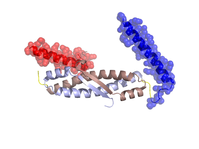
PDBid Chains Hinge Swapped Domain
2OA5 A,B A:41-44,B:41-44 A:6-40,B:6-40

Swapped-domain interface residues and interactions:

Chains Residues
A 18, 21, 22, 25, 28,
B 28, 40, 86,

Non-swapped-domain interface residues and interactions:

Chains Residues
A 41, 42, 43, 44, 48, 49, 50, 52, 53, 54, 56, 57, 60, 61, 64, 68, 69, 72, 73, 76, 79, 80, 81, 82, 83, 85, 86, 89, 90, 92, 93, 94, 95, 96, 97, 98, 99, 101, 106,
B 41, 42, 43, 44, 48, 49, 50, 52, 53, 54, 56, 57, 60, 61, 64, 68, 69, 72, 73, 76, 77, 79, 80, 81, 82, 83, 85, 87, 89, 90, 92, 93, 94, 95, 96, 97, 98, 99, 101,

Swapped domains are represented using trasperent spheres.
Non-swapped part is represented using light color and cartoon representation.
Hinge region is shown in yellow color.

Mutations in critical regions:

Chains Hinge Domain swapped interface Non-swapped interface Swapped Domain
ANo mutationNo mutationNo mutationNo mutation
BNo mutationNo mutationNo mutationNo mutation

HIDE output:

Homologues found through HIDE algorithm

JMOL Visualization:

Jmol_logo            Jmol_logo            Jmol_logo

2D-plot:

A:2OA5 B:2OA5

JOY Structural annotation for hinge hinge and swapped domain:

Hinge Swapped domain

JOY output:

ali file:2OA5.ali
atm file:2OA5.atm
cof file:2OA5.cof
hbd file:2OA5.hbd
html file:2OA5.html
pdb file:2OA5.pdb
ps file:2OA5.ps
psa file:2OA5.psa
rtf file:2OA5.rtf
sst file:2OA5.sst
tem file:2OA5.tem