Pfam Domains mapped on to the structure: 2O2K
No.
Chain ID
Pfam ID
Pfam Description
Linkout - Pfam
Linkout - CDD
1
A
PF02965
Vitamin B12 dependent methionine synthase, activation domain
PF02965
PF02965
Gene Ontology Annotations: 2O2K
Conserved Domain Database Superfamily Annotations: 2O2K
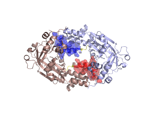
Structural Details of PDB entry 2O2K
Structural Details of PDB entry 2O2K
PDBid Chains Hinge Swapped Domain
2O2K
A,B
A:973-975,B:973-975
A:978-1000,B:978-1000
Swapped-domain interface residues and interactions:
Chains Residues
A
980 , 987 , 988 , 989 , 990 , 991 , 992 , 998 , 1000 , 1071 , 1178 , 1227 , 1228 , 1229 , 1231 ,
B
980 , 987 , 988 , 989 , 990 , 991 , 992 , 998 , 1000 , 1071 , 1167 , 1178 , 1227 , 1228 , 1229 , 1231 ,
Non-swapped-domain interface residues and interactions:
Chains Residues
A
926 , 927 , 976 , 1067 , 1069 , 1070 , 1076 , 1077 , 1079 , 1115 , 1116 , 1117 , 1118 , 1120 , 1165 , 1166 , 1177 , 1226 , 1232 , 1263 ,
B
926 , 927 , 1067 , 1069 , 1070 , 1076 , 1077 , 1079 , 1115 , 1116 , 1117 , 1118 , 1120 , 1165 , 1166 , 1177 , 1226 , 1232 ,
Mutations in critical regions:
Chains
Hinge
Domain swapped interface Non-swapped interface Swapped Domain
A No mutation ASN(1071)LYS, No mutation No mutation B No mutation ASN(1071)LYS, No mutation No mutation
HIDE output:
JMOL Visualization:
2D-plot:
JOY Structural annotation for hinge hinge and swapped domain:
JOY output: