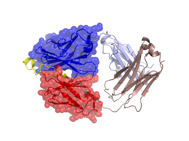
Structural Details of PDB entry 2NY3
Structural Details of PDB entry 2NY3
PDBid Chains Hinge Swapped Domain
2NY3
D,C
D:3102-3117,C:2094-2099
D:3001-3101,C:2001-2093
Swapped-domain interface residues and interactions:
Chains Residues
C
2001 , 2036 , 2038 , 2043 , 2044 , 2046 , 2049 , 2055 , 2056 , 2087 , 2089 , 2091 , 2096 , 2098 , 2100 , 2102 ,
D
3039 , 3043 , 3044 , 3045 , 3047 , 3050 , 3059 , 3062 , 3095 , 3113 , 3114 , 3115 , 3116 , 3118 ,
Non-swapped-domain interface residues and interactions:
Chains Residues
C
2094 , 2095 , 2097 , 2118 , 2120 , 2121 , 2123 , 2125 , 2126 , 2129 , 2131 , 2133 , 2135 , 2137 , 2139 , 2140 , 2162 , 2163 , 2164 , 2165 , 2166 , 2169 , 2176 , 2178 , 2180 , 2182 , 2214 ,
D
3111 , 3119 , 3137 , 3138 , 3139 , 3140 , 3150 , 3152 , 3156 , 3158 , 3179 , 3181 , 3182 , 3184 , 3185 , 3186 , 3194 , 3196 , 3198 , 3224 ,