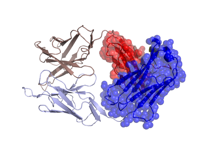
Structural Details of PDB entry 2MPA
Structural Details of PDB entry 2MPA
PDBid Chains Hinge Swapped Domain
2MPA
H,L
H:133-139,L:124-130
H:140-224,L:131-219
Swapped-domain interface residues and interactions:
Chains Residues
H
135 , 136 , 140 , 145 , 149 , 151 , 172 , 173 , 174 , 175 , 177 , 179 , 184 , 186 , 188 , 216 , 221 , 223 ,
L
132 , 136 , 138 , 140 , 142 , 143 , 165 , 166 , 167 , 168 , 169 , 172 , 179 , 181 , 183 , 185 , 191 , 217 , 218 , 219 ,
Non-swapped-domain interface residues and interactions:
Chains Residues
H
35 , 37 , 39 , 44 , 45 , 47 , 59 , 61 , 95 , 99 , 101 , 102 , 107 , 109 , 110 , 111 , 130 , 131 , 132 , 133 , 134 , 138 ,
L
37 , 39 , 41 , 43 , 48 , 49 , 51 , 54 , 55 , 60 , 92 , 96 , 99 , 100 , 101 , 103 , 105 , 119 , 121 , 123 , 124 , 125 , 126 , 128 , 129 , 130 ,