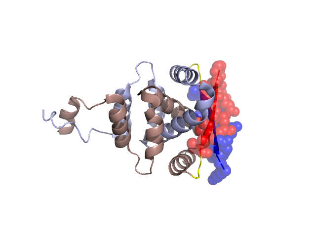
Structural Details of PDB entry 2JD3
Structural Details of PDB entry 2JD3
PDBid Chains Hinge Swapped Domain
2JD3
A,B
A:15-17,B:15-17
A:6-14,B:6-14
Swapped-domain interface residues and interactions:
Chains Residues
A
6 , 7 , 8 , 9 , 10 , 11 , 12 , 13 , 14 , 15 , 16 , 37 , 40 , 41 ,
B
6 , 7 , 8 , 9 , 10 , 11 , 12 , 13 , 14 , 15 , 16 , 37 , 40 , 41 ,
Non-swapped-domain interface residues and interactions:
Chains Residues
A
17 , 18 , 21 , 23 , 24 , 25 , 27 , 28 , 29 , 31 , 39 , 42 , 43 , 44 , 45 , 46 , 47 , 48 , 49 , 50 , 51 , 52 , 53 , 58 , 59 , 61 , 62 , 65 , 69 , 71 , 72 , 74 , 75 , 78 , 82 , 84 , 85 , 86 , 89 , 90 , 91 , 93 , 94 ,
B
17 , 18 , 21 , 23 , 24 , 25 , 27 , 28 , 31 , 39 , 42 , 43 , 44 , 45 , 46 , 47 , 48 , 49 , 50 , 51 , 52 , 53 , 58 , 59 , 61 , 62 , 65 , 69 , 70 , 71 , 72 , 74 , 75 , 78 , 82 , 84 , 85 , 86 , 87 , 89 , 90 , 93 , 95 ,