Home BrowseSearch Help Group

Pfam Domains mapped on to the structure: 2J6G

No. Chain ID Pfam ID Pfam Description Linkout - Pfam Linkout - CDD
1 A PF02432 Fimbrial, major and minor subunit PF02432 PF02432
Structural Details of PDB entry 2J6G

Structural Details of PDB entry 2J6G

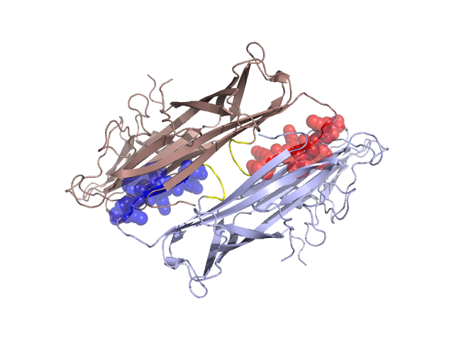
PDBid Chains Hinge Swapped Domain
2J6G A,B A:18-22,B:18-22;A:30-34,B:30-34 A:23-29,B:23-29

Swapped-domain interface residues and interactions:

Chains Residues
A 1, 2, 3, 4, 8, 9, 10, 23, 24, 25, 26, 27, 28, 29, 59, 60, 61, 62, 63, 64, 65, 66, 67,
B 1, 2, 3, 4, 8, 9, 10, 23, 24, 25, 26, 27, 28, 29, 59, 60, 61, 62, 63, 64, 65, 66, 67,

Non-swapped-domain interface residues and interactions:

Chains Residues
A 0, 6, 7, 14, 15, 17, 22, 30, 31, 34, 35, 36, 37, 69, 77, 79, 81, 159, 250, 259, 261, 262,
B 0, 6, 7, 14, 15, 17, 22, 30, 31, 34, 35, 36, 37, 69, 77, 79, 81, 159, 250, 259, 261,

Swapped domains are represented using trasperent spheres.
Non-swapped part is represented using light color and cartoon representation.
Hinge region is shown in yellow color.

Mutations in critical regions:

Chains Hinge Domain swapped interface Non-swapped interface Swapped Domain
ANo mutationNo mutationNo mutationNo mutation
BNo mutationNo mutationNo mutationNo mutation

HIDE output:

Homologues found through HIDE algorithm

JMOL Visualization:

Jmol_logo            Jmol_logo            Jmol_logo

2D-plot:

A:2J6G B:2J6G

JOY Structural annotation for hinge hinge and swapped domain:

Hinge Swapped domain

JOY output:

ali file:2J6G.ali
atm file:2J6G.atm
cof file:2J6G.cof
hbd file:2J6G.hbd
html file:2J6G.html
pdb file:2J6G.pdb
ps file:2J6G.ps
psa file:2J6G.psa
rtf file:2J6G.rtf
sst file:2J6G.sst
tem file:2J6G.tem